
Osteoarthritis Update August 2015 Save

Different Phenotypes for Osteoarthritis of the Foot. Osteoarthritis is often characterized as either a symmetric polyarticular (often involving DIPs, PIPs and CMC1), asymmetric oligoarticular or monarticular (knee or hip OA), but OA may also affect the foot. A recent study sheds light on different phenotypes of OA of the foot – MTP1 OA or more extensive joint involvement that may include the midfoot. (Citation source http://buff.ly/1J8Fch9).
Rathod and coworkers studied 533 adults with foot pain in the prior year. These patients did not have psoriatic or rheumatoid arthritis and had a mean age of 65 years. After collecting data and studying the x-rays, they showed that 15% had polyarticular foot OA, 22% had isolated MTP1 OA, and 64% had minimal or no foot arthritis. Three-quarters of polyarticular patients were women, compared to half with MTP1 OA. Polyarticular foot OA was associated with nodal hand OA, higher body mass index, worse pain scores and disability scores on the Manchester Foot Pain and Disability Index.
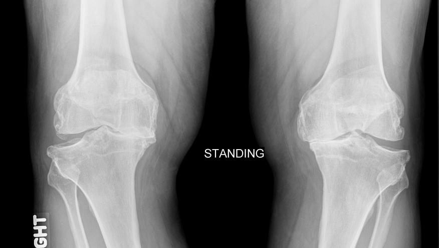
Posttraumatic Total Knee Arthroplasty (TKA) Has Worse Outcomes Compared to TKA for Osteoarthritis. Small studies have shown TKA following a distal femur or tibial fracture have poor outcomes (Citation source http://buff.ly/1PpKINv). This study examined a larger cohort of 531 patients undergoing TKA following post-traumatic fractures and compared their outcomes with 19,641 patients with TKA for osteoarthritis. The periarticular fracture group had significantly revision TKA, infection and complications and worse overall revision free survival compared to with OA, with 1 in 4 patients requiring revision TKA by 15years. A second cohort study revealed the same outcomes with higher complication rates in those undergoing TKA for post-traumatic knee fractures (Citation source http://buff.ly/1gH4Aj3).
Does Severity of Radiographic Osteoarthritis Affect the Outcome? Presumably only the most severe radiographic OA patients become candidates for TKA. However, it is estimated that 1% of TKA recipients have only minimal radiographic change and it is believed that their outcomes may be worse. Perry et al examined a cohort of 29 patients (31 knees) with minimal degenerative changes who underwent TKA and compared their outcomes to a matched cohort with severe radiographic OA. After a mean of 5 yrs (2-10 yrs) both groups showed equal improvement in knees scores, functional and pain measures. Complication rates were higher in the minimal OA group (19%) compared with the severe OA group (3%). Although minimal radiographic OA patients had a higher risk of complications, their pain, functional, and satisfaction outcomes were similar to those with more severe arthritis. (Citation source http://buff.ly/1WoUMLx)
Predictive Value of Low Grade Systemic Inflammation in Symptomatic Knee OA. Attur and colleagues studied a cohort of symptomatic knee OA (SKOA) patients and assessed plasma lipids PGE2 and 15-HETE, and transcriptome analysis of PBLs by microarray and qPCR. They showed that PGE2 and 15-HETE levels were higher in SKOA. PBL transcriptome analysis showed IL-1β, TNFα and COX-2 mRNA in PBLs predicted a greater risk for radiographic progression by joint space narrowing. (Citation source http://buff.ly/1N7E5Ry).
ADD THE FIRST COMMENT
Disclosures
The author has no conflicts of interest to disclose related to this subject


If you are a health practitioner, you may Login/Register to comment.
Due to the nature of these comment forums, only health practitioners are allowed to comment at this time.