

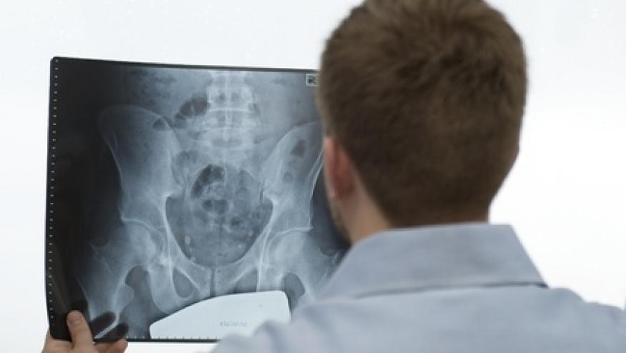
ASAS: MRI Definition of Active Sacroiliitis Save

Increased use of MRI for diagnosis of spondyloarthritis (SpA) in the last two decades has led to earlier diagnoses and gradual incorporation of MRI into the SpA assessment toolset. In 2009, an MRI definition was added to the Assessment in SpondyloArthritis International Society (ASAS) criteria for axial spondyloarthritis. Then the MRI definition was defined as an observation of inflammation in subchondral bone solely, while other MRI findings (fat metaplasia, sclerosis, erosion or ankyloses) were not required.
Investigators with the ASAS MRI working group (16 rheumatologists and 4 radiologists and 1 research fellow) have evaluated the data published in the last 5 years in an effort to better define MRI changes and activity indicative of inflammatory axial SpA. Such modifications would be pivotal to diagnostic and classification considerations.
The ASAS group concluded that the presence of inflammation (defined as Bone Marrow Oedema – BMO) is the principal observation required by the current definition and no additional findings are required.
The description for sacroiliac joint BMO consistent with active SpA is:
- Bone marrow oedema (BMO) on a T2-weighted sequence sensitive for free water (such as short tau inversion recovery (STIR) or T2FS) or bone marrow contrast enhancement on a T1-weighted sequence (such as T1FS post-Gd).
- Inflammation must be clearly present and located in a typical anatomical area (subchondral bone).
- MRI appearance must be highly suggestive of SpA.
Interestingly enough, the sole presence of other inflammatory lesions, such as synovitis, enthesitis or capsulitis, without concomitant BMO is not sufficient for the definition of ‘active sacroiliitis on MRI.
Also, in the absence of MRI signs of BMO, the presence of structural lesions such as fat metaplasia, sclerosis, erosion or ankylosis does not meet the definition of ‘active sacroiliitis on MRI.
The group highlighted following guidelines for MRI interpretation of active sacroiliitis:
- BMO shall be easily seen on at least two consecutive slices of an MRI scan
- The reader of the MRI scan simultaneously review sequences designed to identify inflammation and sequences that focus on depiction of structural damage.
- The presence of concomitant structural damage, especially erosion, and/or other signs of inflammation in the absence of BMO do not suffice to meet the criterion.
The definition and guidelines outlined are meant primarily for the classification of patients with SpA and will not be suitable for use in other clinical situations. See the citation for reference images.
ADD THE FIRST COMMENT
Disclosures
The author has no conflicts of interest to disclose related to this subject


If you are a health practitioner, you may Login/Register to comment.
Due to the nature of these comment forums, only health practitioners are allowed to comment at this time.