Subclinical Heart Inflammation Seen in RA Save

Subclinical myocardial inflammation is common among patients with rheumatoid arthritis (RA) and is associated with articular disease activity, researchers reported.
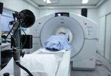
Among a cohort of RA patients without recognized cardiovascular disease who underwent cardiac 18-fluorodeoxyglucose positron emission tomography with computed tomography (FDG PET-CT), 39% had visually assessed FDG uptake and 18% had quantitatively assessed mean standardized uptake values (SUVs) of 3.10 units or higher, according to Joan M. Bathon, MD, of Columbia University in New York City, and colleagues.
In addition, the mean SUV was 31% higher among patients whose Clinical Disease Activity Index (CDAI) was 10 or higher compared with those whose CDAI was less than 10 (P=0.005), the researchers reported online in Arthritis & Rheumatology.
Heart failure is an important factor contributing to cardiovascular disease in RA, and tends to have fewer symptoms but higher mortality than in the wider population. Levels of circulating cytokines such as tumor necrosis factor (TNF) and interleukin-6, which are predictive of heart failure in the general population, are considerably higher in RA, but few studies have addressed inflammation within the myocardium in RA.
Autopsy studies from the mid-20th century identified myocarditis in up to 20% of RA patients, and while the gold standard for the diagnosis of myocarditis is with an endomyocardial biopsy, that test is costly, invasive, and associated with complications.
Today, however, myocardial inflammation also can be detected with FDG PET-CT. "Inflammatory cells are metabolically active and avidly take up FDG via glucose transporters; moreover, areas of myocardial FDG uptake strongly correlate with numbers of infiltrating macrophages and T cells on histologic assessment," Bathon and colleagues explained.
To explore the possibility that inflammation would be detectable in RA patients without apparent heart failure, and to see if this correlated with disease activity, the researchers analyzed 119 patients with RA from the RHYTHM study who had FDG PET-CT scans adequate to assess myocardial FDG uptake.
Participants' mean age was 54, median disease duration was 6.7 years, and most were women. Three-quarters were seropositive, and the majority had low to moderate disease activity.
Most were being treated with disease-modifying antirheumatic drugs, usually methotrexate, and more than one-thirds were on biologics. Prednisone was being used by one-third, and more than 40% were taking nonsteroidal anti-inflammatory drugs.
Troponin-I levels were undetectable in all, indicating absent myocardial damage.
Abnormal FDG uptake was defined as an SUV two standard deviations above the mean of what was seen in a group of 27 healthy controls.
Of the 46 patients with visually assessed myocardial FDG uptake, 25 had only focal uptake and 21 had diffuse uptake. For those with visualized focal uptake, the mean SUV was 61% higher than for those without any visible uptake (2.80 vs 1.74 units, P<0.001), and for those with visualized diffuse myocardial uptake, the mean SUV was 124% higher than those without any uptake (3.89 vs 1.74 units, P<0.001).
On univariate analysis, factors that were associated with higher mean SUV included higher CDAI, higher Disease Activity Score in 28 joints, black race, and higher body mass index, while the use of non-TNF biologics was associated with a lower SUV. After adjustment, only higher disease activity was associated with a higher mean SUV, and non-TNF biologic use (primarily abatacept [Orencia]) was associated with lower mean SUV.
The association between abatacept, which is soluble cytotoxic T-lymphocyte-associated protein 4-immunoglobulin (CTLA4-Ig), and lower myocardial SUV was "tantalizing," the researchers said, pointing out that antibodies that neutralize CTLA4 are used in cancer immunotherapy and have been linked with fatal myocarditis. Nonetheless, they acknowledged that the lower myocardial FDG uptake is probably more strongly associated with decreased RA disease activity than with the specific medication used.
In a substudy that included eight patients with active disease who had escalated therapy after their first FDG PET-CT scan to either TNF inhibitor treatment or triple therapy with methotrexate, sulfasalazine, and hydroxychloroquine, a second scan 6 months later showed that only one patient continued to have visible FDG uptake in the myocardium. The reduction in FDG uptake was similar to the decrease in mean CDAI score.
The finding that disease activity correlated with FDG uptake "supports the premise that achieving low disease activity or remission of RA activity protects not only the joints but possibly the myocardium as well," the investigators observed.
"In summary, although larger longitudinal data are needed, the current study supports the hypotheses that myocardial inflammation in RA is related to disease activity, that it may contribute to the increased risk for heart failure in patients with RA compared with controls, and that it may be responsive to step-up therapy," they concluded.
Limitations of the study, the authors said, included their inability for ethical reasons to provide pathologic verification of the myocardial inflammation and the possibility of other unaccounted variables since the ones examined accounted for only 10% of the variability of observed mean SUV.
The study was supported by the National Institute of Arthritis and Musculoskeletal and Skin Diseases and by the National Institutes of Health National Center for Advancing Translational Sciences.


If you are a health practitioner, you may Login/Register to comment.
Due to the nature of these comment forums, only health practitioners are allowed to comment at this time.