


Transcription
Hi, I'm Dr. Janet Pope. I wanted to present a QD clinic for our gout month at RheumNow. So, this is titled "It is not always RA even when it appears to be."
So, this is a patient starting with this 72-year-old woman from the Philippines. She was assessed with polyarticular inflammatory arthritis. She had lumps on her hands and elbows. Her toes were amputated from the past. She said she had some nodules or lumps that probably got infected and so she had a forefoot amputation on one side.
So just thinking about that, what else should I ask? So I did ask about alcohol, zero. Diet, some fish, no shellfish, no diet soda, not much red meat. Other history, she had onset in her 30s and she's now wheelchair bound at age 72. It started in her hands, elbows, ankles, feet, and spread to her knees, and eventually even her shoulders. She was treated for rheumatoid arthritis elsewhere, and it really didn't help. More recently, she's had hypertension on low-dose hydrochlorothiazide, 12.5 milligrams a day, and she thinks that might have made her problem worse. She was also on aspirin for cardiac protection, 81 milligrams a day, but when she was really having pain and suffering, she was increasing to about six tabs a day. Sometimes a few times a week she would do it, but not always. Family history: interesting. Two brothers, father, and all her uncles on her dad's side had severe gout. No other meds, no allergies.
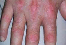
So when I looked at her, she looked her age. She had obvious damage in multiple joints. Her blood pressure was normal. She was in a wheelchair and she did have severe contractures of hands, some MCPs and PIPs, wrists, elbows, and knees. She had the amputation as I described. She had obvious yellow-white lumps in many areas including on the foot at the stump of the amputation, elbows, and elsewhere. She also unfortunately had several swollen joints. It looked like pseudo-rheumatoid arthritis where it was MCPs, PIPs, elbows, even shoulder effusions. One knee was swollen, both knees had flexion contractures, and a DIP was swollen as well.
So, thinking about gout, what are her risk factors? Well, CKD, no, she had a normal creatinine. Diet, no. Lifestyle, no. Meds, only hydrochlorothiazide, long after this started. Family history, strong. Yes, strong. And ethnicity — she's from the Philippines.
And my teaching pearl is often my experience. I can't always validate this, but my experience is patients from the Philippines tend to have mild RA because they're usually not HLA-DR4 positive. They might not have the double shared epitope either, and they tend to have worse gout.
So why? I asked ChatGPT and we did a search — I asked: do patients from the Philippines have a higher prevalence of tophaceous gout and more gout in general? And it's probably mostly a genetic predisposition. The main thing is there are three transporters of uric acid that can be abnormal and more common in patients of Filipino descent. Number one, the ABCG2 urate transporter gene. Number two, the SLC2A9, also called GLUT9. And the third one, SLC22A12, or URAT1. And you've heard of URAT1 transport genes because there are some drugs being developed to help people with gout in this area.
So the variant — especially of the first one I mentioned — the ABCG2 Q141K variant is particularly common in East and Southeast Asian populations. What does this mean? They often have earlier disease onset for gout, in their 30s and 40s. Tophi, joint damage, and chronic erosive changes, and they can also have delayed diagnosis, undertreatment, and misdiagnosis. Diet sometimes might affect this, just like it will in other patient populations. Alcohol is usually not high among my patients anecdotally with gout, particularly the women from the Philippines, and also CKD and hypertension will worsen it. So I think that this is important to be aware of. Am I going to test a genetic variant? No.
Now I have one other brief case. So this is again — all that glitters isn't gold. What is RA by diagnosis might not be. So this is a true case as well. A 54-year-old woman diagnosed with RA a few years ago, referred for a second opinion after failing multiple csDMARDs and a TNF inhibitor, onset in her early 40s, but diagnosis a few years ago. She also had hereditary hemorrhagic telangiectasia, also known as Osler-Rendu syndrome, or HHT. So lots of telangiectasias and often had iron deficiency anemia.
So what did I already know? She had a low-grade positive rheumatoid factor, negative CCP, erosions on X-rays at least by the report description, mild CKD, no other meds, no alcohol, and no dietary red flags. On exam, swollen joints, a slight proximal dactylitis especially the right second finger, ankles, mid feet, had lumps, and the lateral TMT on the fifth part of her foot approximately had a large fluid-filled bursa, lumps at the olecranon bursa, lumps on the Achilles. Working diagnosis was: is this RA or gout or both? So, I
did the natural thing aspirating the lump on her right um uh TMT midfoot area and uh uh what came out would actually looked like milk. And when we sent it to the lab knowing it'd be filled with crystals, um they first wouldn't analyze it said, "Are you uh um doing an April Fool's joke?" Because it was in early April uh because it looks like milk. And we said, "No, analyze it." And for sure it was filled with tophi.
So I asked the family history. Her son had gout onset in his early 20s. Um, and sure enough, he is now my patient too with tophaceous gout, no alcohol, and he's on treatment. And my patient was adopted. So, family history of her parents, etc. was unknown.
So, let's fast forward. She's been on high-dose allopurinol after eight years. Now, all tophi are gone. And she's very thankful and thinks I'm a genius. I'm not a genius. We just had to do the history physical and think about it.
So learning tips from this case and the other um history and family history are really important clues. History and physical will give clues to mimics of RA. So pseudo RA but there should be DIP involved and it might be asymmetrical and the lumps under the skin are often white or yellow whereas rheumatoid nodules might be slightly yellow. They're really never white. um the distribution more lower than upper extremity, feet greater than hands, and location, location, location. Go for where the lumps are, where the joints are. And if you're not doing the right thing, do a uric acid and obviously put a needle in aspirated joint. I think failure of other treatment, especially if they're diagnosed with another form of RA, re-examine the X-rays or do them. And if in doubt, as I say, get a needle, pull out uh fluid and get it analyzed for negatively birefringent crystals. Thank you. I hope you enjoyed these cases.
So, this is a patient starting with this 72-year-old woman from the Philippines. She was assessed with polyarticular inflammatory arthritis. She had lumps on her hands and elbows. Her toes were amputated from the past. She said she had some nodules or lumps that probably got infected and so she had a forefoot amputation on one side.
So just thinking about that, what else should I ask? So I did ask about alcohol, zero. Diet, some fish, no shellfish, no diet soda, not much red meat. Other history, she had onset in her 30s and she's now wheelchair bound at age 72. It started in her hands, elbows, ankles, feet, and spread to her knees, and eventually even her shoulders. She was treated for rheumatoid arthritis elsewhere, and it really didn't help. More recently, she's had hypertension on low-dose hydrochlorothiazide, 12.5 milligrams a day, and she thinks that might have made her problem worse. She was also on aspirin for cardiac protection, 81 milligrams a day, but when she was really having pain and suffering, she was increasing to about six tabs a day. Sometimes a few times a week she would do it, but not always. Family history: interesting. Two brothers, father, and all her uncles on her dad's side had severe gout. No other meds, no allergies.
So when I looked at her, she looked her age. She had obvious damage in multiple joints. Her blood pressure was normal. She was in a wheelchair and she did have severe contractures of hands, some MCPs and PIPs, wrists, elbows, and knees. She had the amputation as I described. She had obvious yellow-white lumps in many areas including on the foot at the stump of the amputation, elbows, and elsewhere. She also unfortunately had several swollen joints. It looked like pseudo-rheumatoid arthritis where it was MCPs, PIPs, elbows, even shoulder effusions. One knee was swollen, both knees had flexion contractures, and a DIP was swollen as well.
So, thinking about gout, what are her risk factors? Well, CKD, no, she had a normal creatinine. Diet, no. Lifestyle, no. Meds, only hydrochlorothiazide, long after this started. Family history, strong. Yes, strong. And ethnicity — she's from the Philippines.
And my teaching pearl is often my experience. I can't always validate this, but my experience is patients from the Philippines tend to have mild RA because they're usually not HLA-DR4 positive. They might not have the double shared epitope either, and they tend to have worse gout.
So why? I asked ChatGPT and we did a search — I asked: do patients from the Philippines have a higher prevalence of tophaceous gout and more gout in general? And it's probably mostly a genetic predisposition. The main thing is there are three transporters of uric acid that can be abnormal and more common in patients of Filipino descent. Number one, the ABCG2 urate transporter gene. Number two, the SLC2A9, also called GLUT9. And the third one, SLC22A12, or URAT1. And you've heard of URAT1 transport genes because there are some drugs being developed to help people with gout in this area.
So the variant — especially of the first one I mentioned — the ABCG2 Q141K variant is particularly common in East and Southeast Asian populations. What does this mean? They often have earlier disease onset for gout, in their 30s and 40s. Tophi, joint damage, and chronic erosive changes, and they can also have delayed diagnosis, undertreatment, and misdiagnosis. Diet sometimes might affect this, just like it will in other patient populations. Alcohol is usually not high among my patients anecdotally with gout, particularly the women from the Philippines, and also CKD and hypertension will worsen it. So I think that this is important to be aware of. Am I going to test a genetic variant? No.
Now I have one other brief case. So this is again — all that glitters isn't gold. What is RA by diagnosis might not be. So this is a true case as well. A 54-year-old woman diagnosed with RA a few years ago, referred for a second opinion after failing multiple csDMARDs and a TNF inhibitor, onset in her early 40s, but diagnosis a few years ago. She also had hereditary hemorrhagic telangiectasia, also known as Osler-Rendu syndrome, or HHT. So lots of telangiectasias and often had iron deficiency anemia.
So what did I already know? She had a low-grade positive rheumatoid factor, negative CCP, erosions on X-rays at least by the report description, mild CKD, no other meds, no alcohol, and no dietary red flags. On exam, swollen joints, a slight proximal dactylitis especially the right second finger, ankles, mid feet, had lumps, and the lateral TMT on the fifth part of her foot approximately had a large fluid-filled bursa, lumps at the olecranon bursa, lumps on the Achilles. Working diagnosis was: is this RA or gout or both? So, I
did the natural thing aspirating the lump on her right um uh TMT midfoot area and uh uh what came out would actually looked like milk. And when we sent it to the lab knowing it'd be filled with crystals, um they first wouldn't analyze it said, "Are you uh um doing an April Fool's joke?" Because it was in early April uh because it looks like milk. And we said, "No, analyze it." And for sure it was filled with tophi.
So I asked the family history. Her son had gout onset in his early 20s. Um, and sure enough, he is now my patient too with tophaceous gout, no alcohol, and he's on treatment. And my patient was adopted. So, family history of her parents, etc. was unknown.
So, let's fast forward. She's been on high-dose allopurinol after eight years. Now, all tophi are gone. And she's very thankful and thinks I'm a genius. I'm not a genius. We just had to do the history physical and think about it.
So learning tips from this case and the other um history and family history are really important clues. History and physical will give clues to mimics of RA. So pseudo RA but there should be DIP involved and it might be asymmetrical and the lumps under the skin are often white or yellow whereas rheumatoid nodules might be slightly yellow. They're really never white. um the distribution more lower than upper extremity, feet greater than hands, and location, location, location. Go for where the lumps are, where the joints are. And if you're not doing the right thing, do a uric acid and obviously put a needle in aspirated joint. I think failure of other treatment, especially if they're diagnosed with another form of RA, re-examine the X-rays or do them. And if in doubt, as I say, get a needle, pull out uh fluid and get it analyzed for negatively birefringent crystals. Thank you. I hope you enjoyed these cases.




If you are a health practitioner, you may Login/Register to comment.
Due to the nature of these comment forums, only health practitioners are allowed to comment at this time.