
All News

Keys to Mastery (5.2.2025)
Dr. Jack Cush reviews the news, articles and drug approvals from the past week on RheumNow.com. This podcast marks the beginning of our Lupus Campaign called "Lupus Unlocked: Keys to Mastery". This month's campaign on Lupus is sponsored by Aurinia.
Read Article
Osteoarthritis Risky Business (4.18.2025)
Dr. Jack Cush reviews the news and Journal reports from this week on RheumNow.com. Osteoarthritis patients have unique risks and synovial fluid WBC numbers can tell you when to worry about septic arthritis in gout and pseudogout patients.
Read Article
EMR Messaging Woes (4.11.2025)
Dr. Jack Cush reviews the news, journal reports and regulatory approvals from this past week on RheumNow.com
Read Article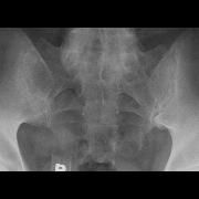
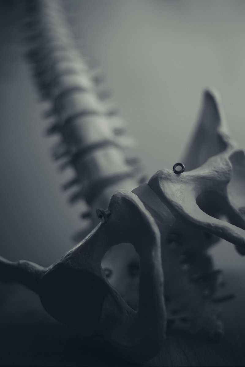
Pitfalls in sacroiliitis imaging: Bone marrow edema may also be seen in young-middle-aged postpartum women, & athletes & kids (ongoing bone growth) & w/ advancing age (DJD) https://t.co/bwS047wlmI https://t.co/FvWlEcZ5JP
Dr. John Cush RheumNow ( View Tweet)
Rheumatologist Survey: What do you rely on to diagnose neuropsychiatric lupus (cerebritis)?
Dr. John Cush RheumNow ( View Tweet)
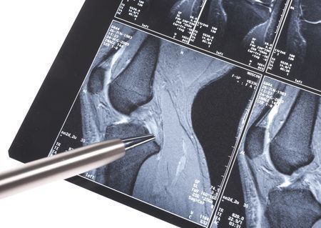
Osteoarthritis Initiative (OAI) longitudinal study analyzed 1075 #OA knees (1/pt) in 96 mo. MRI study to predict outcomes. Infrapatellar fat pad (IPFP volume & signal) predicted future knee replacement & long-term damage. IPFP correlated w/ cartil. volume & BM lesions; Not Sxs. https://t.co/Xsju7JdEVE
Dr. John Cush RheumNow ( View Tweet)
RHEUMATOLOGIST Survey: What are your contraindications tor renal Bx?
Dr. John Cush RheumNow ( View Tweet)
RHEUMATOLOGIST Survey: What are your contraindications tor renal Bx?
Dr. John Cush RheumNow ( View Tweet)

Osteoarthritis Initiative (OAI) longitudinal study analyzed 1075 #OA knees (1/pt) in 96 mo. MRI study to predict outcomes. Infrapatellar fat pad (IPFP volume & signal) predicted future knee replacement & long-term damage. IPFP correlated w/ cartil. volume & BM lesions; Not https://t.co/VrLfiSGWGA
Dr. John Cush RheumNow ( View Tweet)
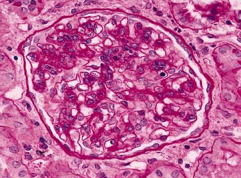
Simplistic overview of Lupus Nephritis - causes, risk factors, symptoms, diagnosis, and treatment of lupus nephritis. https://t.co/QkLXUQcSqo https://t.co/pGjCHmoelG
Dr. John Cush RheumNow ( View Tweet)
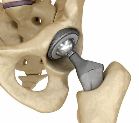
Hip replacement outcomes are not linked to OA severity, instead pts muscle quality before THA best predicts recovery after surgery. Imaging of 10 pts undergoing THA showed poor muscle quality predicted post-op performance/movement after surgery https://t.co/io0UXQl6n2 https://t.co/cFM7Fg10Zm
Dr. John Cush RheumNow ( View Tweet)

Hip replacement outcomes are not linked to OA severity, instead pts muscle quality before THA best predicts recovery after surgery. Imaging of 10 pts undergoing THA showed poor muscle quality predicted post-op performance/movement after surgery https://t.co/io0UXQl6n2 https://t.co/eI7u6BTPYx
Dr. John Cush RheumNow ( View Tweet)
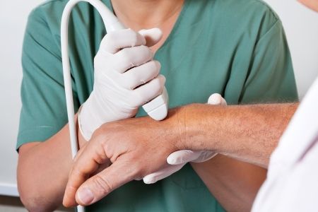
Ultrasound lesions of dactylitis associ w/short-term development of PsA. Study of 64 PsO +arthralgia pts. IUS PD synovitis & B mode enthesitis predicted PsA Dx (p < .001) w/ an accuracy of 89.1 %, a sensitivity of 86.7 % and a specificity of 89.8 %. https://t.co/KoXMmoxXhP https://t.co/tj2NxKDkqh
Links:
Dr. John Cush RheumNow ( View Tweet)
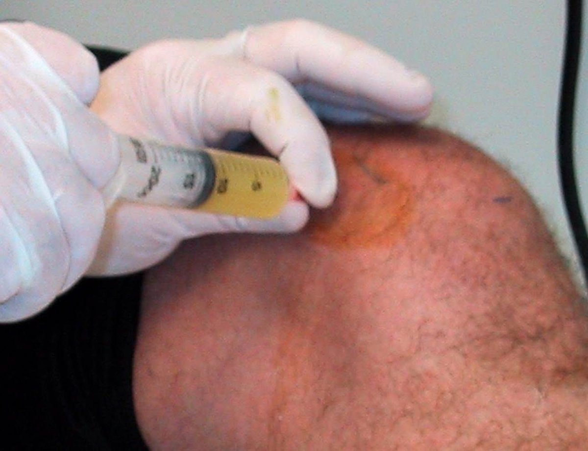
Distinguishing Septic and Gouty Arthritis
A single center, retrospective review of patients undergoing knee joint fluid aspirations for presumed crystalline arthritis (CA) showed that synovial WBC may provide a useful diagnostic marker for SA with an optimal threshold of 50,000 https://t.co/ye69mRooSh
Dr. John Cush RheumNow ( View Tweet)
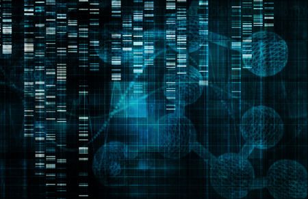
Genomics and Future Drug Targets for Osteoarthritis
A Nature article details the results of an osteoarthritis, genome-wide association study meta-analyses across up to 489,975 cases and 1,472,094 controls, establishing 962 independent associations, 513 of which have not been https://t.co/OYyePkAeAM
Dr. John Cush RheumNow ( View Tweet)

Ultrasound lesions of dactylitis associ w/short-term development of PsA. Study of 64 PsO +arthralgia pts. IUS PD synovitis & B mode enthesitis predicted PsA Dx (p < .001) w/ an accuracy of 89.1 %, a sensitivity of 86.7 % and a specificity of 89.8 %. https://t.co/Ij6oYP1TuK https://t.co/55sO4uFXNI
Links:
Dr. John Cush RheumNow ( View Tweet)
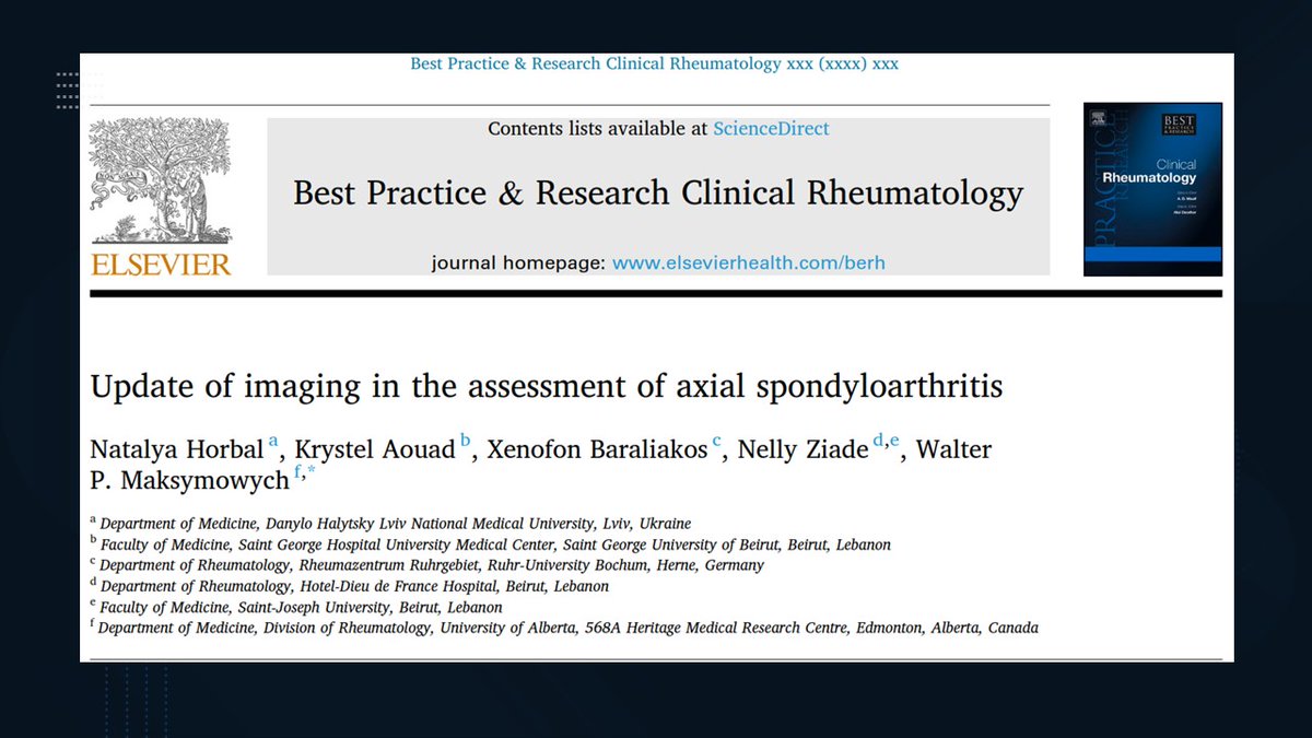
🚨 New publication alert!
We review the latest advances in imaging for axial spondyloarthritis—from high-res MRI and synthetic CT to deep learning algorithms improving lesion detection.
🔗 https://t.co/oddjoFOGzM
#AxSpA #AIinMedicine
@XBaraliakos @WalterMaks @krystelaouad https://t.co/VwuUwYL1Xs
Links:
Nelly ZIADE 🍀 Nellziade ( View Tweet)
KORAIL (Korean RA-ILD) prospective cohort study of CT and biomarkers showed 35% ILD progressed over 3 yrs. Higher baseline KL-6 (HR 1.37) & hSP-D (1.51) levels assoc w/ progression. Increasing KL-6 at 1 yr showed the greatest progression (∆KL-6: HR 2.0) https://t.co/efowyCp1qu https://t.co/JVd7sRKzVF
Dr. John Cush RheumNow ( View Tweet)
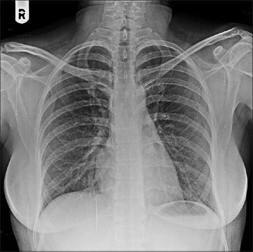
Diagnostically, Xrays are effective & sufficient at Diagnosing chronic low back pain; X-rays predicted MRI results in approximately 75% of the cases for common low back pain problems suggesting they may be a preferred initial imaging study for this condition. https://t.co/PYzd1zXIRz
Dr. John Cush RheumNow ( View Tweet)
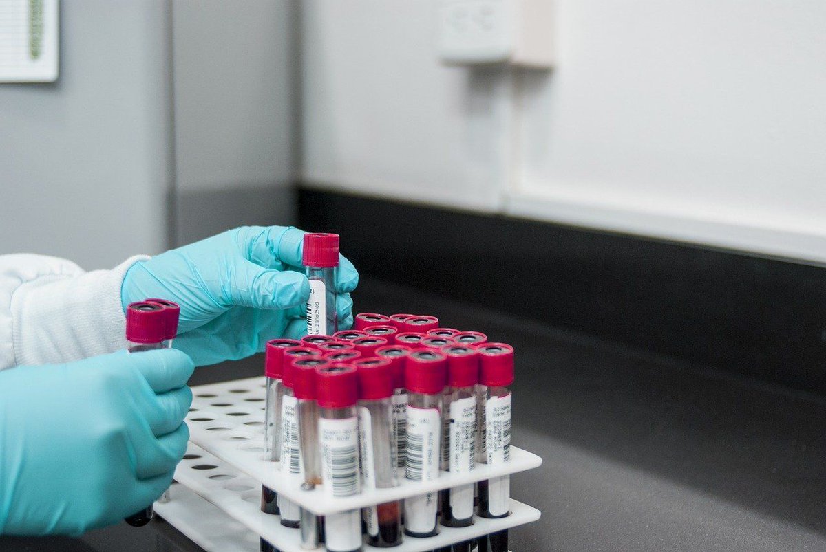
Consensus on Bone Turnover Biomarkers in Osteoporosis
A new consensus paper, published by the European Society for Clinical and Economic Aspects of Osteoporosis, Osteoarthritis and Musculoskeletal Diseases (ESCEO), the International Osteoporosis Foundation (IOF), and the https://t.co/lq1odNBLdZ
Dr. John Cush RheumNow ( View Tweet)

