Axial Spondylitis Abstracts Roundup Save

There have been a number of interesting posters and presentations covering the field of axial spondyloarthropathy. I’ve picked out a few points that I found particularly relevant.
Classification Criteria
Attending the study group on axSpA, I was reassured to note that the ASAS classification criteria are going to be studied again. There are large international studies underway of new presentation of ‘inflammatory back pain’ (before HLA B27 is known) - SPARTAN in the US, CLASSIC worldwide. They have pre-specified that to be adopted the criteria must show at least 90% specificity and 75% sensitivity. I think that a number of clinicians (myself included) are concerned that the nr-axSpA group seems to have a different gender and genetic make up and not all cases progress to ‘AS’ as we know it. The CLASSIC study will include central reading of MRI scans, which is not as straightforward as we might have thought.
Imaging
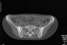
There were a lot of presentations dealing with how to interpret the imaging of the SI joints and spinal areas.
Abstract 897: Dr Baraliakos reported the results of a study involving MRI of the spine and SI joints in 802 young healthy people - 18% had bone marrow oedema in the SI joints and fatty lesions were seen in 86%. Specificity was a little better if the lesions were thoracic and if there were more than 5 lesions in total.
Abstract 1194: Dr Aydin (Ottowa) presented a poster showing the results of a study where ultrasound was performed at 5 sites in 80 healthy subjects (using an ultrasound machine with excellent resolution and doppler sensitivity). She found that 10% had a positive doppler signal, while 6% had erosions. Positive findings in this healthy group was associated with age, BMI, male sex and physical activity. Several other posters mentioned that the ‘enthesophyte’ is very common especially at the calcaneum and should not be used a maker or enthesitis.
Abstract 1206: Dr Verhoeven (France) has scanned anterior chest wall joints in 136 patients with spondyloarthropathy. The incidence of positive findings in the manubristernal and sternoclavicular joints rose from 37% to 63% over a period of 5 years. Only half of the patients were complaining of anterior chest wall pain, so I think that this area will be a useful one to check back in my ultrasound clinic.
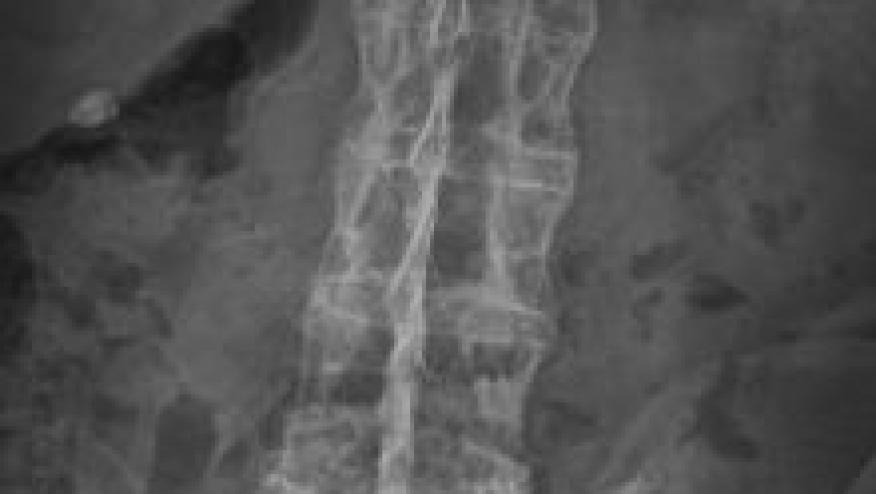
Abstract 1620: Dr Fitzgerald (Ireland) presented a study comparing lateral DXA with AP DXA in diagnosing osteoporosis in 110 axSpA patients with paired lateral and PA DXAs. The lateral spine BMD was significantly lower than AP BMD and detected more osteoporosis (16% vs 2%). Using all sites, 52% of the cohort was designated as having a low BMD compared to 36% when the AP results were used. I knew that it was difficult to measure BMD accurately in axSpA, and this study has helped to bring some clarity. Unfortunately not all DXA scanners report the lateral results.
Early Diagnosis of axSpA (average delay in most countries 7-8 years)
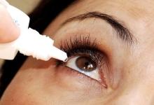
A common theme in many posters this year was an awareness that anterior uveitis occurs in 10-26% of patients with axSpA (less common in PsA and nr-axSpA). This might just be of academic interest, but it actually provides an important opportunity to diagnose axSpA early. In abstract 1639 Dr Lopez-Medina (Cordoba, Spain) used the REGISPONSER registry to examine this in more detail. Her group found that the diagnosis of uveitis predated the diagnosis of axSpA in 36%, an average of 5 years earlier. Since the HLA B27 test was positive in 80% of these patients it gives our Ophthalmology colleagues a great opportunity to help us reduce the long delay in diagnosing axSpA.
Looking at it another way, about a third of patients with acute anterior uveitis will turn out to have axSpA, especially if they also have a history of back pain (A1646). So we need to have a conversation with the ophthalmologists. And if they do us a favour we can do one in return: 22% of patients develop complications from their first episode of uveitis - so if we educate our patients with axSpA about the symptoms and signs they should present earlier to our ophthalmology colleagues.
Another way to make an early diagnosis might be to use a genetic test. It is clear that the positive predictive value of a positive HLA-B27 on its own is low (2-5%), but there could be a potential of using other genetic markers. Dr Li (Plenary abstract 836) presented the results of applying a polygenic risk score using over 1000 SNPs. She reported that the PPV was 15% for the genetic score. I’m sure we will be hearing more about polygenic risk scores in future. She also pointed out that at $50 for the test, it is cheaper than either an MRI or an HLA-B27 test.
Abstract 648 reported the ‘OptiRef’ self-report questionnaire for early axSpA diagnosis. I think most of us would recoil at the idea of a self report diagnosis, but 19% of those self-referring using the tool had a final diagnosis of axSpA compared to 40% of those referred from their primary care physician. Another poster showed that using advanced physiotherapists as an intermediary step also improved early diagnosis.
I hope you’ve found some useful information here that is relevant to your practice.


If you are a health practitioner, you may Login/Register to comment.
Due to the nature of these comment forums, only health practitioners are allowed to comment at this time.