Chikungunya Club Meeting (7.24.2026)
Dr. Jack Cush reviews the news, journal reports from the past week on RheumNow.com.
Read Article
Search for Terms or Answers

Dr. Jack Cush reviews the news, journal reports from the past week on RheumNow.com.
Read Article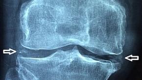
A recent perspective by Tedeschi tackles many unresolved issues around CPPD diagnosis, classification, and management, including the lack of clinical trials or FDA approved treatments.
Read Article

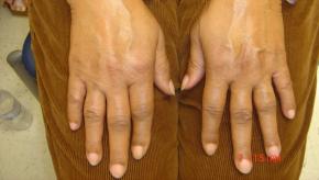
Years of IL-17 vs. IL-23 debate in PsA finally got tested head-to-head: bimekizumab outperformed risankizumab on joint outcomes in the BE BOLD trial, while a GLP-1 combination study suggests weight and metabolic status are active drivers of PsA — not just comorbidities. This recap also digs into
Read Article
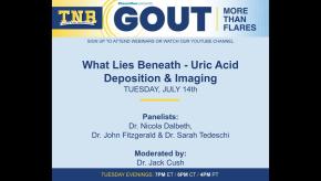
Dr. Jack Cush reviews the news, journal articles and Gout features from this past week on RheumNow.com.
Read Article


International guidelines can help identify dermatomyositis patients with a particularly high risk for cancer, according to a retrospective single-center U.S. study.
Read Article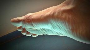
Gout is all about the crystals and the consequences of the disease are due to the inflammation they cause. Getting rid of urate crystals is the ultimate goal in gout, as we now know that it leads to the risk zero of flares and it normalizes the cardiovascular risk level of patients
Read Article
Dr. Jack Cush reviews the news, journal articles and regulatory decisions from the past week. July is Gout: More than Flares month.
Read Article
In a recent JAMA Viewpoints article, Dr. Susan Ott addresses several controversies in preventing osteoporosis (OP) related fractures.
Read Article

A network meta-analysis in the Journal of Autoimmunity reviews the pharmacologic options for rheumatoid arthritis–associated interstitial lung disease (RA-ILD). RA-ILD guidelines were recently presented by ERS/EULAR, but these were mainly conditional, low-certainty recommendations.
Read Article
Almost everyone will deal with back pain at some point in their lives. Most recover quickly — but for about 20% of people, acute pain becomes a chronic condition that interferes with daily life and keeps them out of the workforce
Read Article
People with rheumatoid arthritis are at risk of developing interstitial lung disease (RA-ILD), which is associated with high mortality. ANCHOR-RA is a large, international cross-sectional prospective study that will enable the development of a multivariable model to help detect RA-ILD.5
Read Article
Dr. Jack Cush reviews the news and journal articles from RheumNow.com. Updates on tofacitinib, CAR-T therapy and AI.
Read Article
Biomarker interest has grown considerably in the last 2 decades, owing to advances in genetics, imaging, protein, and multiomics. Despite these advances, biomarkers as the predictive holy grail of RA therapeutics and prognostication have not yet advanced beyond rheumatoid serologies and C-
Read Article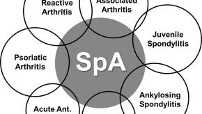
An editorial in Lancet Rheumatology calls for "..reframing axSpA as a systemic, autoimmune inflammatory disease with multiorgan involvement and substantial unmet need."
The pathogenesis relies on not only HLA-B27, but also on environmental triggers, immune dysregulation, and
Read Article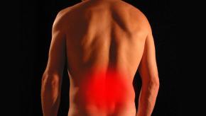
A comprehensive JAMA review synthesizes current evidence on the epidemiology, pathophysiology, clinical evaluation, and treatment of nonspecific low back pain, drawing on 108 publications identified from a PubMed search (2005–2026) and the most recent guidelines from the WHO, ACP, and NICE.
Read Article
On the final day of EULAR 2026, Mukhtyar et al (on behalf of a large international task force) presented the updated EULAR recommendations for management of polymyalgia rheumatica (PMR), giant cell arteritis (GCA), and Takayasu arteritis (TAK). There were 5 overarching principles and 12
Read Article
As EULAR2026 comes to a close, practical learnings take precedence as clinicians head back to their clinics. Among them, the 2026 EULAR Update on Imaging Recommendations in SpA stands out.
Read Article

Imaging in the preclinical phase of RA is moving fast—arguably faster than our ability to interpret what we are actually seeing. Across EULAR 2026 abstracts, a consistent theme emerges: we are improving detection of subclinical inflammation, but still struggling to determine what
Read Article


By downloading this material, I acknowledge that it may be used only for personal use and personal education and that I will accredit RheumNow.com as the source and owner of this material. Commercial use or mass reproduction of this material without permission from RheumNow (info@rheumnow.com) is prohibited.