Imaging and Early Detection in Psoriatic Arthritis Save
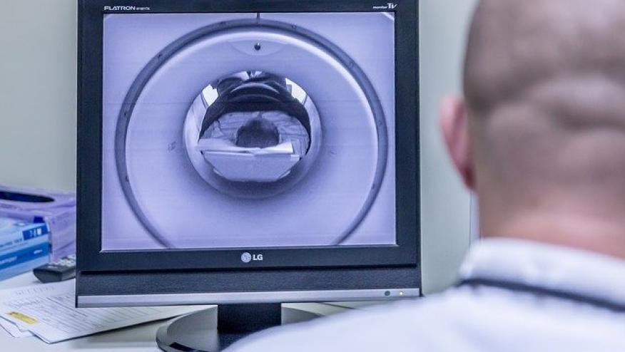
At EULAR 2025, there have been new developments in imaging in PsA. Two key poster presentations (POS0104 and OP0179) provide important insights into the role of imaging in the management and early detection of psoriatic arthritis (PsA).
On the poster tour, the TIGERS study (POS0104) was presented. This study explored the use of whole-body MRI (WB-MRI) to monitor treatment response in PsA patients receiving different biologic therapies. In this randomized controlled trial, 32 PsA patients were assigned to receive adalimumab (TNF inhibitor), guselkumab (IL-23p19 inhibitor), or ustekinumab (IL-23 inhibitor). Imaging was performed using WB-MRI with the MRI-WIPE scoring system, and standard clinical assessments were collected, including TJC68, SJC66, DAPSA, and Leeds Enthesitis Index. The results demonstrated that adalimumab led to a significant reduction in MRI-WIPE scores (median decrease of 39 units) and joint synovitis scores (median decrease of 23 units), while guselkumab and ustekinumab did not show significant imaging changes. All three treatments improved clinical scores, but only adalimumab achieved significant reductions in both imaging and clinical measures. There was a strong correlation between MRI synovitis and SJC66 changes (rho 0.78, p=0.023), suggesting that WB-MRI can capture inflammation not fully reflected in clinical scores. These findings highlight the potential of WB-MRI as a valuable tool to assess treatment response and personalise therapy in PsA, particularly for identifying subclinical inflammation.
In parallel, the RAPSODI and PSOART cohorts (OP0179) investigated the role of subclinical inflammation in the early development of PsA among psoriasis patients. In this prospective study, 226 psoriasis patients underwent clinical and ultrasound assessments to detect subclinical joint or enthesis involvement. Over a median follow-up of 12 months, 31 individuals developed PsA, with most presenting peripheral arthritis, predominantly oligoarthritis. Importantly, 68% of these patients had evidence of subclinical abnormalities at the anatomical sites that later became clinically involved. Specifically, 26% had tenderness alone, 32% had ultrasound-detected abnormalities, and 42% had both. Ultrasound most commonly revealed low-grade synovitis (grade 1-2) in small joints and wrists, with only a single case of entheseal involvement. These data suggest that ultrasound can identify joints at risk for progression to clinically manifest PsA, supporting the concept of "very early PsA" and emphasizing the importance of targeted monitoring in psoriasis patients.
Together, these studies underscore the evolving role of advanced imaging modalities in PsA management.
WB-MRI offers a comprehensive assessment of inflammatory burden that can guide therapeutic decisions and detect residual inflammation despite clinical remission. Ultrasound, meanwhile, may serve as an early predictor of joints likely to develop PsA, potentially enabling earlier diagnosis and intervention to modify disease progression. The integration of these imaging techniques into routine PsA care could lead to more precise and personalised treatment approaches.





If you are a health practitioner, you may Login/Register to comment.
Due to the nature of these comment forums, only health practitioners are allowed to comment at this time.