

APR: Systemic Lupus Erythematosus Save
Transcription
This is Advanced Practice RheumNow. Hi, I'm Jack Cush, RheumNow. In this review, we'll be discussing systemic lupus and what you need to know and understand to manage this complex disorder.
Lupus is sort of the prototypic autoimmune disorder where it's really a disorder of adaptive immunity. It's a chronic inflammatory disease where there's a loss of self-tolerance and there's the development of autoantibodies and immune complexes that give rise to the many manifestations which are so typical. So lupus is an autoimmune disease characterized by a systemic presentation, multi-organ involvement, multiple autoantibodies and immune complex deposition. The etiology of the disorder is unknown. This is a strongly female predominant disorder 10 to one female to male and the genetics are important but even in monozygotic twins there's only a 30% concordance rate.
Lupus is not as big as everyone thinks it is because most people misdiagnose it based on the presence of an ANA which is found in up to 20 million Americans. 15% of people who are sick or elderly, but it's really only seen in about 300,000 or less in the United States. The peak onset is between 15 and 40 years of age, but usually the average age of a person being included in a lupus trial might be in the late 40s or early 50s. As I said, it's female to male 10 to one. But that female predominance is lost when you get into the elderly age groups.
In this disorder especially, there is a racial disparity that affects not only the risk of getting the disease, but also manifesting more severe disease. Lupus is more severe and these groups are also at higher risk if you're African, Asian, Hispanic, Latin, also in children and males. It tends to be milder in Caucasians, elderly, and what is called drug-induced lupus.
The diagnosis of lupus — there are new EULAR/ACR criteria for classification. You must have a number of points. There's a point system. It's really quite complex and really not memorable. These came out in 2018 and you can look them up or put them on your phone. I like to remember the 1987 ACR criteria for lupus where you needed to have four out of 11 criteria because I can remember these. And you start out by remembering four skin — there's four skin manifestations: malar rash, painless oral ulcers, photosensitivity, and discoid rash. It's rheumatology so arthritis is number five. It's lupus so there's ANAs and other autoantibodies — that's six and seven — ANAs and an immunologic criteria which is either a double-stranded DNA, Sm antibody, or an antiphospholipid antibody, or biologic false positive VDRL or RPR. So the other ones that remain that you have to remember are renal — and that's having nephrotic syndrome or RBC casts. Next, serositis, and lastly CNS manifestations, either seizures or psychosis. So in total there's 11; you need to have four at any time to then be considered for diagnosis. You need to have a number of criteria to make ordering the ANA of any value. We'll get to that in a second.
So lupus manifests as sort of mild, moderate, and severe disease. Mild lupus, very common in Caucasians or the onset of disease, maybe elderly, is constitutional, cutaneous, and musculoskeletal manifestations. Those are better managed with simple drugs like non-steroidals, antimalarials. More serious manifestations like serositis, nephritis, neuropsychiatric disease, and hematologic disease require higher dose steroids, disease-modifying and immunosuppressant therapies and maybe biologics. And then renal disease and problematic refractory disease will require newer therapies, and then we have problems of cardiopulmonary disease and complications of disease.
The main thing you need to know from a clinical standpoint is the skin. The malar rash is the most distinctive, highly specific feature of lupus. It is on the face. It's over the malar prominences, and it really shows up on sun-exposed areas. So top of forehead, the malar ridge, maybe upper lip, and maybe chin. It spares the nasolabial fold. This is incredibly important because a rash that goes over a nasolabial fold is not the malar rash of lupus. A rash that goes over the nasolabial fold could be a number of other things, mainly rosacea. But there are other things like lupus pernio and periorbital cellulitis and polymorphous light eruption — photosensitive skin eruptions. So I have a few pictures of that in the slide handout for you to look at.
When characterizing lupus skin disease, there's really three types. And you can characterize lupus skin disease based on the type of rash. There's three types: the distribution, the associated symptoms, systemic or not, and the autoantibody profile. The worst is acute LE skin disease. These are people with autoantibodies — double-stranded DNA and Sm antibodies in addition to ANA. All three types of lupus skin disease will have ANAs. Thus, the ANA is not that helpful. But
specific double-strand DNA SM is more systemic disease, acute manifestations in acute LE rash is the malar rash, the oral ulcers, photosensitivity, alopecia and bullous eruptions and those are people with high amount of systemic manifestations. The mildest is called chronic cutaneous and we also call that discoid LE where there's scarring and alopecia. They're going to be ANA positive but really no other distinctive auto-antibody profile and no other systemic features and they have the best prognosis and then in between is what's called subacute LE or SCLE and there's two forms — annular forms would sort of round like rashes and papulosquamous rashes that look like psoriasis they're called psoriasiform — these people have some systemic manifestations and tend to be unified with an auto-antibody against SSA or Ro antibodies. So that's how you understand lupus skin disease.
The other important skin manifestation of lupus is Raynaud's. How do you diagnose Raynaud's? It's not red, white, and blue — the colors of the American flag. It is first white — loss of all blood supply due to arterial constriction. That's the first and sometimes missed finding of Raynaud's. And then when the artery is clenched down, you have pooling of venous blood and then you get this dusky cyanosis or the blue appearance which is next most common and then when the person warms up the skin there's a hyperreactive erythema and the red manifestation. So the order here is white then blue then red. You need to have at least two phases or biphasic features to really qualify as Raynaud's.
You see Raynaud's in lupus but also in the two different forms of systemic sclerosis — diffuse systemic sclerosis and then limited systemic sclerosis also called CREST. It's also seen in MCTD. It's a characteristic feature and in polymyositis and dermatomyositis, much less in other disorders like RA and Sjögren's.
Musculoskeletal features. Arthralgia is very common and not very specific. Arthritis less common, more specific — likes to be in PIPs, polyarticular PIPs, knees and then maybe others. Erosive severe inflammatory arthritis is quite uncommon but you can get deformities and they're reducible deformities and that's called Jaccoud's arthritis. Lupus patients can also get joint pain from fibromyalgia. That's called type 2 lupus. They get joint pain from being on steroids, having osteoporosis and developing fractures or osteonecrosis. Very uncommon is myositis or overlap features.
The ANA is paramount to the diagnosis. We have another advanced practice rheumatology video and teaching session on the ANA and auto-antibodies. Look at that. Having an ANA is not synonymous with a diagnosis of lupus. It's overly sensitive and poorly specific. You want specific — look for double-stranded DNA and SM antibodies. You can find ANAs in many conditions: drug induced, being elderly, being chronically ill, chronic liver disease, chronic renal disease, chronic lung disease, neoplasia, pregnancy. It's a lousy screen for arthritis. I don't care if you're ANA positive. That is not a diagnosis. I need other features to make the diagnosis.
So the rules on ordering ANAs: don't order an ANA unless two or more lupus features or criteria are evident. Hence the ANA is only useful in the presence of symptoms that are specific for lupus. Don't repeat ANAs — one and done is all you need. Don't order a sub-serology like an ENA or double-stranded DNA if the ANA is negative; it has very little yield. And I don't think that repeating ANAs of 1:40 or 1:80 are needed, but you certainly can. Most of those are not going to be lupus. ANA-negative lupus does not exist.
The SM antibody and double-stranded DNA are found in 20 to 50% of patients and can be really very specific. The other important antibody in lupus is SSA antibodies also called Ro antibodies. The associations are multiple. SSA antibodies are found in Sjögren's syndrome — SS meaning Sjögren's syndrome associated A — but it's also found in lupus, subacute cutaneous lupus, that's that in-between version of lupus skin disease. Neonatal lupus syndrome with heart block and C2 deficiency and rashes in neonates. Elder onset lupus is often SSA positive. SSA can be associated with C2 and C4 deficiencies and also primary biliary cirrhosis. Prior to 1985, all ANA-negative lupus was really due to SSA antibodies.
You need to look at other labs. The CBC is very important. Leukopenia is common in lupus and I like to see white counts less than 3,000 although you qualify as leukopenia at a white count of less than 4,000. Lymphopenia less than 1,500 absolute lymphocytes — you qualify; less than 1,100 is scary. Lymphopenia can be due to active lupus. It can be due to the immunosuppressant you're using to treat lupus such as azathioprine, mycophenolate. And then thrombocytopenia — again under 400,000 you start to qualify, but really under 150 or 100,000 is a worrisome feature of lupus. Much less common is anemia but it really
to be lupus related it has to be a hemolytic anemia associated with an autoantibody and then lastly the antiphospholipid syndrome. Antiphospholipid syndrome as you know is the triad of having either thrombotic events or recurrent spontaneous abortions or thrombocytopenia in the presence of an antiphospholipid antibody manifest as either an anticardiolipin antibody, a biologic false positive RPR — no syphilis, just RPR — and that's due to an antiphospholipid antibody, or having a prolonged PTT that doesn't correct with serum. This is also called a lupus anticoagulant and there are multiple tests that you can do for that. I like the dRVVT, the dilute Russell Viper Venom Time, or the platelet neutralization test, and there are multiples.
There are other features that can go along with antiphospholipid antibodies that are part of the syndrome although they're not the major criteria. We're talking about migraine headaches, livedo reticularis, sterile endocarditis, what's called Libman-Sacks endocarditis, a mitral regurgitation, transverse myelitis, and even unexplained neuropathies.
These antiphospholipid antibodies by themselves are not pathologic. They're found in multiple autoimmune disorders and connective tissue disorders. They're found in about 30% of lupus patients, but 30% of lupus patients don't have the antiphospholipid syndrome. You have to have the antibody plus the classic presentation. So the disease antiphospholipid syndrome correlates not with the antibody but also the co-occurrence of antibodies against beta-2 glycoprotein 1. When present you might treat these people with anticoagulants such as warfarin, heparin, or dax rather than immunosuppressants.
The antiphospholipid syndrome again you should probably be looking for in someone who has a diagnosis of lupus. An important clinical feature of lupus is coronary artery disease because it contributes to mortality. There's a bimodal pattern of death in lupus. Early deaths in lupus are due to the disease and infection. Later deaths after age 50 are predominantly due to cardiovascular disease and it is a major cause of morbidity and mortality in lupus, found in somewhere between six and 10% of patients.
We worry most about the occurrence of lupus nephritis because this also has a strong correlation with bad outcomes. You classify the patient according to the WHO based on the renal biopsy. Type one is a normal biopsy. Everybody with lupus has a type two or worse. Type two is mesangial proliferative. Type three is focal proliferative where less than 50% of the glomeruli are affected. Type four or worse than type three is diffuse proliferative where more than 50% of the glomeruli are affected. And that's 50% of the number of glomeruli found on the biopsy. You find 11 glomeruli, you've got to have six or more to be affected to have class 4 diffuse proliferative. Type five is membranous glomerulonephritis which gives you a lot of proteinuria, usually nephrotic range proteinuria, and then a really bad finding — or not part of the classification system — is interstitial disease, interstitial fibrosis. Patients are scored as far as their activity index and their chronicity index. Again, the things that are really specific for lupus and bad lupus are proliferative lesions, positive immunofluorescence, subendothelial deposits, and a high activity index more so than chronicity index.
How does lupus nephritis present? Most of it is silent and many academic centers will biopsy patients without symptoms looking for nephritis. But you worry about nephritis in the presence of microscopic hematuria with or without casts, the presence of hypertension, new onset renal insufficiency, new onset hypertension, and then proteinuria might be suspected based on the urinalysis but also peripheral edema, periorbital edema, or anasarca.
A worrisome feature is neuropsychiatric lupus. What we used to call lupus cerebritis makes the presentation as either diffuse disease, seizures, psychosis, or cognitive dysfunction. But even organic brain syndrome and headache is included as diffuse disease. It can have more focal presentations such as strokes, transverse myelitis — those two could be related to antiphospholipid syndrome — cranial or peripheral neuropathies, ataxia, and movement disorders.
The frequency of neuropsychiatric disease if you really look at it is about 50%. We don't know why it happens. There is no diagnostic test. You have to have a high index of suspicion because there is no gold standard. The diagnosis is often made in retrospect and you really need to do a combination of things. One, neuropsychiatric lupus has no correlation with lupus activity. So really you have to do an LP and do cell counts, protein, glucose, cultures for fungi, mycobacteria, regular bacteria, a VDRL, and cryptococcal antigen. You want to see if there's inflammation and immunologic activity within the brain tissue by looking at the CSF IgG and albumin production. Order a Q albumin,
order the IgG index, order anti-neuronal antibodies. You do CNS imaging mainly to rule out other causes or mass problems within the brain — MRI or CT are just fine — and then there's talk of functional measures, but I don't and I've never found utility in doing neuropsychiatric or cognitive testing, EEGs, or evoked potentials, but some advocate for those.
The pearls on neuropsychiatric disease are the following. One: a lupus patient admitted to the hospital with brain manifestations is more likely to have neuropsychiatric lupus than a CNS infection or steroid psychosis, and that's the exact opposite of a lupus patient who gets admitted to the hospital with another problem — a lung problem, a blood problem, a lupus activity problem — without brain disease. They're more likely to be admitted to the hospital for a non-lupus reason more so than a lupus reason. So again, with brain manifestations you really have to think neuropsychiatric.
It will present in people who have well-established lupus. It is very very very very rarely the first manifestation of lupus. It has no concordance with active clinical lupus or serologic lupus or even autopsy findings. The onset can occur while they're on lupus meds, off lupus meds, and I'm talking strong meds like immunosuppressants, cytotoxics, and biologics.
Don't overlook the mild symptoms. Cognitive dysfunction is a major issue, and the worst headache they've ever had — think lupus serositis or neuropsychiatric lupus.
The main findings in a CSF is a lymphocytic pleocytosis. WBC counts are low, you know 2 to 10, 2 to 20, with a lymphocytic predominance — not a poly predominance, lymphocytic — and they may or may not have high protein, but those two findings are only seen in probably 20–30% of patients with neuropsychiatric lupus. If you want a higher yield from the CSF, meaning greater than 50% suggestion of cerebritis, you've got to order those CSF tests of Q albumin, IgG index, IgG synthetic rate, and oligoclonal bands.
Lupus CNS lupus is the second leading cause of death. Occurs in 10 to 25% of deaths. And if they have it early, it means they're going to have a bad course for their lupus. The occurrence of transverse myelitis is also correlated with poor outcomes, and that, as I said earlier, is associated with antiphospholipid antibodies.
Two more main points. Complications of lupus include, one, overlap syndromes. RA and lupus overlap is called Rhupus. MCTD and undifferentiated connective tissue disease is really an overlap where you have overlaps of lupus with either myositis, scleroderma, or RA.
Infection is the number one cause of death, and you get this in lupus because of immunosuppression, chronic and high-dose steroids, and then maybe complement deficiencies.
Osteonecrosis is a big complication, especially with chronic steroid use. One joint, especially a large joint — shoulder, hip, knee — think osteonecrosis. Get an X-ray or MRI.
Alveolar hemorrhage. Wow. When this happens, it's bad news. They may present with hemoptysis, shortness of breath, a cough that's not productive. They have a sudden drop in PO2 and a whiteout on their chest X-ray due to the alveoli being filled with blood. The differential diagnosis for this presentation — acute onset, often with fever — is either infectious pneumonia versus lupus pneumonitis versus alveolar hemorrhage. These people should be hospitalized and worked up.
Vasculitis, although it's common throughout rheumatology, is actually quite rare in lupus and usually only small vessel, usually presenting as either palpable purpura or leukocytoclastic vasculitis.
There is, as I said earlier, excess mortality from cardiovascular disease and atherosclerotic disease.
Libman-Sacks endocarditis is antiphospholipid antibody associated, and the neonatal lupus syndrome — children born to SSA or Ro-positive mothers who are born with congenital heart block, rash, and a C2 deficiency.
There's a slide in the handout looking at the management of lupus where you manage based on the severity of disease. Mild disease — skin, constitutional, joint, plus or minus serositis — sunscreen, topicals, prednisone, and non-steroidals might be all you need. The next level would be refractory arthritis, refractory skin disease, hematologic disease — may require methotrexate, TNF inhibitors, steroids, hydroxychloroquine. In fact, everybody with lupus should be on hydroxychloroquine; it is vitamin H.
Severe disease — nephritis, CNS disease, severe hematologic, alveolar hemorrhage — you're talking about high-dose steroids, immunosuppressants, azathioprine, mycophenolate, cyclophosphamide, newer agents: belimumab, voclosporin, anifrolumab, and obinutuzumab. Obinutuzumab and voclosporin are only approved for lupus nephritis.
And then if you have the APS syndrome, you're talking about anticoagulation with heparin, warfarin, and maybe DOACs.
One last important teaching point: hydroxychloroquine. It's probably the safest drug we use in rheumatology. It's probably the safest drug you'll ever use in managing lupus. And everyone should be
on it, no matter what. If they're going to have chronic lupus, they should be on hydroxychloroquine. Why? Being on hydroxychloroquine has six major benefits. It decreases the frequency of neonatal lupus and cardiac complications. Second, it lowers fasting glucose, improves insulin resistance and lowers the rate of associated diabetes with lupus. Three, it decreases the risk of thrombotic events. Being on hydroxychloroquine is basically an anticoagulant of sorts. Four, it improves survival in multiple studies. It decreases flare rates in lupus. And then lastly, adherence to hydroxychloroquine overall lowers lupus mortality.
That's it for this edition of Advanced Practice Rheum. Tune in for more.
Lupus is sort of the prototypic autoimmune disorder where it's really a disorder of adaptive immunity. It's a chronic inflammatory disease where there's a loss of self-tolerance and there's the development of autoantibodies and immune complexes that give rise to the many manifestations which are so typical. So lupus is an autoimmune disease characterized by a systemic presentation, multi-organ involvement, multiple autoantibodies and immune complex deposition. The etiology of the disorder is unknown. This is a strongly female predominant disorder 10 to one female to male and the genetics are important but even in monozygotic twins there's only a 30% concordance rate.
Lupus is not as big as everyone thinks it is because most people misdiagnose it based on the presence of an ANA which is found in up to 20 million Americans. 15% of people who are sick or elderly, but it's really only seen in about 300,000 or less in the United States. The peak onset is between 15 and 40 years of age, but usually the average age of a person being included in a lupus trial might be in the late 40s or early 50s. As I said, it's female to male 10 to one. But that female predominance is lost when you get into the elderly age groups.
In this disorder especially, there is a racial disparity that affects not only the risk of getting the disease, but also manifesting more severe disease. Lupus is more severe and these groups are also at higher risk if you're African, Asian, Hispanic, Latin, also in children and males. It tends to be milder in Caucasians, elderly, and what is called drug-induced lupus.
The diagnosis of lupus — there are new EULAR/ACR criteria for classification. You must have a number of points. There's a point system. It's really quite complex and really not memorable. These came out in 2018 and you can look them up or put them on your phone. I like to remember the 1987 ACR criteria for lupus where you needed to have four out of 11 criteria because I can remember these. And you start out by remembering four skin — there's four skin manifestations: malar rash, painless oral ulcers, photosensitivity, and discoid rash. It's rheumatology so arthritis is number five. It's lupus so there's ANAs and other autoantibodies — that's six and seven — ANAs and an immunologic criteria which is either a double-stranded DNA, Sm antibody, or an antiphospholipid antibody, or biologic false positive VDRL or RPR. So the other ones that remain that you have to remember are renal — and that's having nephrotic syndrome or RBC casts. Next, serositis, and lastly CNS manifestations, either seizures or psychosis. So in total there's 11; you need to have four at any time to then be considered for diagnosis. You need to have a number of criteria to make ordering the ANA of any value. We'll get to that in a second.
So lupus manifests as sort of mild, moderate, and severe disease. Mild lupus, very common in Caucasians or the onset of disease, maybe elderly, is constitutional, cutaneous, and musculoskeletal manifestations. Those are better managed with simple drugs like non-steroidals, antimalarials. More serious manifestations like serositis, nephritis, neuropsychiatric disease, and hematologic disease require higher dose steroids, disease-modifying and immunosuppressant therapies and maybe biologics. And then renal disease and problematic refractory disease will require newer therapies, and then we have problems of cardiopulmonary disease and complications of disease.
The main thing you need to know from a clinical standpoint is the skin. The malar rash is the most distinctive, highly specific feature of lupus. It is on the face. It's over the malar prominences, and it really shows up on sun-exposed areas. So top of forehead, the malar ridge, maybe upper lip, and maybe chin. It spares the nasolabial fold. This is incredibly important because a rash that goes over a nasolabial fold is not the malar rash of lupus. A rash that goes over the nasolabial fold could be a number of other things, mainly rosacea. But there are other things like lupus pernio and periorbital cellulitis and polymorphous light eruption — photosensitive skin eruptions. So I have a few pictures of that in the slide handout for you to look at.
When characterizing lupus skin disease, there's really three types. And you can characterize lupus skin disease based on the type of rash. There's three types: the distribution, the associated symptoms, systemic or not, and the autoantibody profile. The worst is acute LE skin disease. These are people with autoantibodies — double-stranded DNA and Sm antibodies in addition to ANA. All three types of lupus skin disease will have ANAs. Thus, the ANA is not that helpful. But
specific double-strand DNA SM is more systemic disease, acute manifestations in acute LE rash is the malar rash, the oral ulcers, photosensitivity, alopecia and bullous eruptions and those are people with high amount of systemic manifestations. The mildest is called chronic cutaneous and we also call that discoid LE where there's scarring and alopecia. They're going to be ANA positive but really no other distinctive auto-antibody profile and no other systemic features and they have the best prognosis and then in between is what's called subacute LE or SCLE and there's two forms — annular forms would sort of round like rashes and papulosquamous rashes that look like psoriasis they're called psoriasiform — these people have some systemic manifestations and tend to be unified with an auto-antibody against SSA or Ro antibodies. So that's how you understand lupus skin disease.
The other important skin manifestation of lupus is Raynaud's. How do you diagnose Raynaud's? It's not red, white, and blue — the colors of the American flag. It is first white — loss of all blood supply due to arterial constriction. That's the first and sometimes missed finding of Raynaud's. And then when the artery is clenched down, you have pooling of venous blood and then you get this dusky cyanosis or the blue appearance which is next most common and then when the person warms up the skin there's a hyperreactive erythema and the red manifestation. So the order here is white then blue then red. You need to have at least two phases or biphasic features to really qualify as Raynaud's.
You see Raynaud's in lupus but also in the two different forms of systemic sclerosis — diffuse systemic sclerosis and then limited systemic sclerosis also called CREST. It's also seen in MCTD. It's a characteristic feature and in polymyositis and dermatomyositis, much less in other disorders like RA and Sjögren's.
Musculoskeletal features. Arthralgia is very common and not very specific. Arthritis less common, more specific — likes to be in PIPs, polyarticular PIPs, knees and then maybe others. Erosive severe inflammatory arthritis is quite uncommon but you can get deformities and they're reducible deformities and that's called Jaccoud's arthritis. Lupus patients can also get joint pain from fibromyalgia. That's called type 2 lupus. They get joint pain from being on steroids, having osteoporosis and developing fractures or osteonecrosis. Very uncommon is myositis or overlap features.
The ANA is paramount to the diagnosis. We have another advanced practice rheumatology video and teaching session on the ANA and auto-antibodies. Look at that. Having an ANA is not synonymous with a diagnosis of lupus. It's overly sensitive and poorly specific. You want specific — look for double-stranded DNA and SM antibodies. You can find ANAs in many conditions: drug induced, being elderly, being chronically ill, chronic liver disease, chronic renal disease, chronic lung disease, neoplasia, pregnancy. It's a lousy screen for arthritis. I don't care if you're ANA positive. That is not a diagnosis. I need other features to make the diagnosis.
So the rules on ordering ANAs: don't order an ANA unless two or more lupus features or criteria are evident. Hence the ANA is only useful in the presence of symptoms that are specific for lupus. Don't repeat ANAs — one and done is all you need. Don't order a sub-serology like an ENA or double-stranded DNA if the ANA is negative; it has very little yield. And I don't think that repeating ANAs of 1:40 or 1:80 are needed, but you certainly can. Most of those are not going to be lupus. ANA-negative lupus does not exist.
The SM antibody and double-stranded DNA are found in 20 to 50% of patients and can be really very specific. The other important antibody in lupus is SSA antibodies also called Ro antibodies. The associations are multiple. SSA antibodies are found in Sjögren's syndrome — SS meaning Sjögren's syndrome associated A — but it's also found in lupus, subacute cutaneous lupus, that's that in-between version of lupus skin disease. Neonatal lupus syndrome with heart block and C2 deficiency and rashes in neonates. Elder onset lupus is often SSA positive. SSA can be associated with C2 and C4 deficiencies and also primary biliary cirrhosis. Prior to 1985, all ANA-negative lupus was really due to SSA antibodies.
You need to look at other labs. The CBC is very important. Leukopenia is common in lupus and I like to see white counts less than 3,000 although you qualify as leukopenia at a white count of less than 4,000. Lymphopenia less than 1,500 absolute lymphocytes — you qualify; less than 1,100 is scary. Lymphopenia can be due to active lupus. It can be due to the immunosuppressant you're using to treat lupus such as azathioprine, mycophenolate. And then thrombocytopenia — again under 400,000 you start to qualify, but really under 150 or 100,000 is a worrisome feature of lupus. Much less common is anemia but it really
to be lupus related it has to be a hemolytic anemia associated with an autoantibody and then lastly the antiphospholipid syndrome. Antiphospholipid syndrome as you know is the triad of having either thrombotic events or recurrent spontaneous abortions or thrombocytopenia in the presence of an antiphospholipid antibody manifest as either an anticardiolipin antibody, a biologic false positive RPR — no syphilis, just RPR — and that's due to an antiphospholipid antibody, or having a prolonged PTT that doesn't correct with serum. This is also called a lupus anticoagulant and there are multiple tests that you can do for that. I like the dRVVT, the dilute Russell Viper Venom Time, or the platelet neutralization test, and there are multiples.
There are other features that can go along with antiphospholipid antibodies that are part of the syndrome although they're not the major criteria. We're talking about migraine headaches, livedo reticularis, sterile endocarditis, what's called Libman-Sacks endocarditis, a mitral regurgitation, transverse myelitis, and even unexplained neuropathies.
These antiphospholipid antibodies by themselves are not pathologic. They're found in multiple autoimmune disorders and connective tissue disorders. They're found in about 30% of lupus patients, but 30% of lupus patients don't have the antiphospholipid syndrome. You have to have the antibody plus the classic presentation. So the disease antiphospholipid syndrome correlates not with the antibody but also the co-occurrence of antibodies against beta-2 glycoprotein 1. When present you might treat these people with anticoagulants such as warfarin, heparin, or dax rather than immunosuppressants.
The antiphospholipid syndrome again you should probably be looking for in someone who has a diagnosis of lupus. An important clinical feature of lupus is coronary artery disease because it contributes to mortality. There's a bimodal pattern of death in lupus. Early deaths in lupus are due to the disease and infection. Later deaths after age 50 are predominantly due to cardiovascular disease and it is a major cause of morbidity and mortality in lupus, found in somewhere between six and 10% of patients.
We worry most about the occurrence of lupus nephritis because this also has a strong correlation with bad outcomes. You classify the patient according to the WHO based on the renal biopsy. Type one is a normal biopsy. Everybody with lupus has a type two or worse. Type two is mesangial proliferative. Type three is focal proliferative where less than 50% of the glomeruli are affected. Type four or worse than type three is diffuse proliferative where more than 50% of the glomeruli are affected. And that's 50% of the number of glomeruli found on the biopsy. You find 11 glomeruli, you've got to have six or more to be affected to have class 4 diffuse proliferative. Type five is membranous glomerulonephritis which gives you a lot of proteinuria, usually nephrotic range proteinuria, and then a really bad finding — or not part of the classification system — is interstitial disease, interstitial fibrosis. Patients are scored as far as their activity index and their chronicity index. Again, the things that are really specific for lupus and bad lupus are proliferative lesions, positive immunofluorescence, subendothelial deposits, and a high activity index more so than chronicity index.
How does lupus nephritis present? Most of it is silent and many academic centers will biopsy patients without symptoms looking for nephritis. But you worry about nephritis in the presence of microscopic hematuria with or without casts, the presence of hypertension, new onset renal insufficiency, new onset hypertension, and then proteinuria might be suspected based on the urinalysis but also peripheral edema, periorbital edema, or anasarca.
A worrisome feature is neuropsychiatric lupus. What we used to call lupus cerebritis makes the presentation as either diffuse disease, seizures, psychosis, or cognitive dysfunction. But even organic brain syndrome and headache is included as diffuse disease. It can have more focal presentations such as strokes, transverse myelitis — those two could be related to antiphospholipid syndrome — cranial or peripheral neuropathies, ataxia, and movement disorders.
The frequency of neuropsychiatric disease if you really look at it is about 50%. We don't know why it happens. There is no diagnostic test. You have to have a high index of suspicion because there is no gold standard. The diagnosis is often made in retrospect and you really need to do a combination of things. One, neuropsychiatric lupus has no correlation with lupus activity. So really you have to do an LP and do cell counts, protein, glucose, cultures for fungi, mycobacteria, regular bacteria, a VDRL, and cryptococcal antigen. You want to see if there's inflammation and immunologic activity within the brain tissue by looking at the CSF IgG and albumin production. Order a Q albumin,
order the IgG index, order anti-neuronal antibodies. You do CNS imaging mainly to rule out other causes or mass problems within the brain — MRI or CT are just fine — and then there's talk of functional measures, but I don't and I've never found utility in doing neuropsychiatric or cognitive testing, EEGs, or evoked potentials, but some advocate for those.
The pearls on neuropsychiatric disease are the following. One: a lupus patient admitted to the hospital with brain manifestations is more likely to have neuropsychiatric lupus than a CNS infection or steroid psychosis, and that's the exact opposite of a lupus patient who gets admitted to the hospital with another problem — a lung problem, a blood problem, a lupus activity problem — without brain disease. They're more likely to be admitted to the hospital for a non-lupus reason more so than a lupus reason. So again, with brain manifestations you really have to think neuropsychiatric.
It will present in people who have well-established lupus. It is very very very very rarely the first manifestation of lupus. It has no concordance with active clinical lupus or serologic lupus or even autopsy findings. The onset can occur while they're on lupus meds, off lupus meds, and I'm talking strong meds like immunosuppressants, cytotoxics, and biologics.
Don't overlook the mild symptoms. Cognitive dysfunction is a major issue, and the worst headache they've ever had — think lupus serositis or neuropsychiatric lupus.
The main findings in a CSF is a lymphocytic pleocytosis. WBC counts are low, you know 2 to 10, 2 to 20, with a lymphocytic predominance — not a poly predominance, lymphocytic — and they may or may not have high protein, but those two findings are only seen in probably 20–30% of patients with neuropsychiatric lupus. If you want a higher yield from the CSF, meaning greater than 50% suggestion of cerebritis, you've got to order those CSF tests of Q albumin, IgG index, IgG synthetic rate, and oligoclonal bands.
Lupus CNS lupus is the second leading cause of death. Occurs in 10 to 25% of deaths. And if they have it early, it means they're going to have a bad course for their lupus. The occurrence of transverse myelitis is also correlated with poor outcomes, and that, as I said earlier, is associated with antiphospholipid antibodies.
Two more main points. Complications of lupus include, one, overlap syndromes. RA and lupus overlap is called Rhupus. MCTD and undifferentiated connective tissue disease is really an overlap where you have overlaps of lupus with either myositis, scleroderma, or RA.
Infection is the number one cause of death, and you get this in lupus because of immunosuppression, chronic and high-dose steroids, and then maybe complement deficiencies.
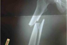
Osteonecrosis is a big complication, especially with chronic steroid use. One joint, especially a large joint — shoulder, hip, knee — think osteonecrosis. Get an X-ray or MRI.
Alveolar hemorrhage. Wow. When this happens, it's bad news. They may present with hemoptysis, shortness of breath, a cough that's not productive. They have a sudden drop in PO2 and a whiteout on their chest X-ray due to the alveoli being filled with blood. The differential diagnosis for this presentation — acute onset, often with fever — is either infectious pneumonia versus lupus pneumonitis versus alveolar hemorrhage. These people should be hospitalized and worked up.
Vasculitis, although it's common throughout rheumatology, is actually quite rare in lupus and usually only small vessel, usually presenting as either palpable purpura or leukocytoclastic vasculitis.
There is, as I said earlier, excess mortality from cardiovascular disease and atherosclerotic disease.
Libman-Sacks endocarditis is antiphospholipid antibody associated, and the neonatal lupus syndrome — children born to SSA or Ro-positive mothers who are born with congenital heart block, rash, and a C2 deficiency.
There's a slide in the handout looking at the management of lupus where you manage based on the severity of disease. Mild disease — skin, constitutional, joint, plus or minus serositis — sunscreen, topicals, prednisone, and non-steroidals might be all you need. The next level would be refractory arthritis, refractory skin disease, hematologic disease — may require methotrexate, TNF inhibitors, steroids, hydroxychloroquine. In fact, everybody with lupus should be on hydroxychloroquine; it is vitamin H.
Severe disease — nephritis, CNS disease, severe hematologic, alveolar hemorrhage — you're talking about high-dose steroids, immunosuppressants, azathioprine, mycophenolate, cyclophosphamide, newer agents: belimumab, voclosporin, anifrolumab, and obinutuzumab. Obinutuzumab and voclosporin are only approved for lupus nephritis.
And then if you have the APS syndrome, you're talking about anticoagulation with heparin, warfarin, and maybe DOACs.
One last important teaching point: hydroxychloroquine. It's probably the safest drug we use in rheumatology. It's probably the safest drug you'll ever use in managing lupus. And everyone should be
on it, no matter what. If they're going to have chronic lupus, they should be on hydroxychloroquine. Why? Being on hydroxychloroquine has six major benefits. It decreases the frequency of neonatal lupus and cardiac complications. Second, it lowers fasting glucose, improves insulin resistance and lowers the rate of associated diabetes with lupus. Three, it decreases the risk of thrombotic events. Being on hydroxychloroquine is basically an anticoagulant of sorts. Four, it improves survival in multiple studies. It decreases flare rates in lupus. And then lastly, adherence to hydroxychloroquine overall lowers lupus mortality.
That's it for this edition of Advanced Practice Rheum. Tune in for more.
Join The Discussion
Dr. Cush, would you really have us go back 40 years–to SLE criteria that had 83% sensitivity (less for atypical disease), while 2019 EULAR/ACR criteria have 96% sensitivity–just because you have a bad memory? You don't have to remember the point system. Use the SLE App.
A paradigm shift has happened in SLE since 1987. Please don't drag the diagnostic process back to a time when early disease and late-onset disease were missed and no one knew any better. This is STILL happening because clinical practitioners are clinging to outdated criteria they were trained on. This leads to misdiagnosis, missed diagnosis, and delayed diagnosis–ultimately leading to diagnostic failure and untreated disease progression for patients. This is not progress.
Late-onset SLE (diagnosed after age 50; comprising up to 20% of SLE patients) barely gets a mention here. The phenotype is well-recognized and should be highlighted for new practitioners: 1) more prevalent in Caucasians; 2) fewer classic skin symptoms; 3) less remarkable antibody profiles; 4) higher prevalence of aPL; 5) more serositis; and 6) lower disease activity, despite higher morbidity and organ damage. Use 2019 criteria to identify it!


If you are a health practitioner, you may Login/Register to comment.
Due to the nature of these comment forums, only health practitioners are allowed to comment at this time.