Best Imaging in Giant Cell Arteritis - US, PET, MRI? Save
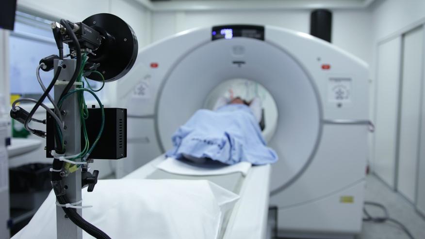
Imaging is often instrumental in diagnosing and staging patients diagnosed with giant cell arteritis (GCA). A new study compared the diagnostic performance of Colour Duplex Ultrasound (CDUS), Fluor-18-deoxyglucose Positron Emission Tomography Computed Tomography (FDG-PET/CT) and Magnetic Resonance Imaging (MRI) in patients suspected of giant cell arteritis (GCA) and found the CDUS had a numerically higher sensitivity (that was not statitically superior to other modalities).
A nested-case control pilot study compared CDUS, whole body FDG-PET/CT and cranial MRI in 23 patients with GCA and 19 patients suspected of but not diagnosed with GCA. Imaging was performed within 5 working days after initial clinical evaluation.
Results are shown in the table below:
Sensitivity | Specificity | |
| CDUS | 69.6% (95%CI 50.4%-88.8%) | 100% |
| FDG-PET | 52.2% (95%CI 31.4%-73.0%) | 100% |
| MRI | 56.5% (95%CI 35.8%-77.2%) | 100% |
FDG-PET/CT was negative for GCA in those with isolated cranial GCA patients (n = 8) and MRI was negative in all isolated extracranial GCA patients (n = 4).
In 4 GCA patients with false-negative results required further imaging confirmed diagnosis.
While CDUS had a higher sensitivity, the confidence intervals of all imaging modalities were overlapping.
EULAR recommendations suggest that CDUS can be used as a first test to diagnose GCA.





If you are a health practitioner, you may Login/Register to comment.
Due to the nature of these comment forums, only health practitioners are allowed to comment at this time.