

Imaging in Axial Spondyloarthritis Save

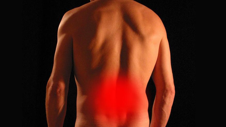
The diagnosis of axial spondyloarthritis (axSpA) is clinical, however, there are still unmet needs, particularly in the diagnosis of early disease and evaluation of disease activity and progression.
Abstract OP0307, a South Korean study, aimed to improve the clinical diagnosis of axSpA by utilizing a deep learning model that integrated structural and inflammatory changes on sacrum MRI, combining data from BME with STIR and T1-weighted images. Data from 291 patients with chronic back pain who underwent MRI were analyzed. A total of 163 patients were classified as having axSpA, with a sensitivity of 87.5%, specificity of 84.0%, and accuracy of 85.96%. Interestingly the model was also able to classify a few axSpA patients who met clinical but not imaging criteria according to ASAS. While this study highlights the application of artificial intelligence in predicting clinical diagnosis, in real-world practice there is a need for a universally accepted algorithm.
In terms of disease progression, abstract OP0310 utilized data from the SPACE cohort to compare structural lesions on the spine in early axSpA vs non-axSpA patients with chronic back pain. Patients underwent conventional radiography (CR) and MRI at baseline and at 2 years. A total of 278 patients (61% HLAB27 positive) underwent both imaging modalities. Over 2 years, there were no differences in the progression of structural changes between both groups in CR, however in MRI, there was a significant increase in the number of fat lesions observed in axSpA patients compared to non-axSpA patients where no lesions were seen, raising the importance of fat lesions as a potential marker of disease progression even in early disease.
Given the evidence presented above, the question of whether there’s still a role for CR in the initial diagnosis of axSpA was raised. This was explored in abstract OP0313 where the diagnostic efficacy of CR, MRI, or Low-dose CT (LDCT) of the SIJ region was evaluated in patients with suspected axSpA. A total of 176 patients with suspected axSpA from a center in Germany were divided into 3 groups, the standard imaging arm underwent all three imaging modalities (XR followed by MRI followed by LDCT if all previous were negative), the MRI arm underwent MRI followed by LDCT if the previous was negative and the LDCT arm underwent LDCT if MRI was negative. In the standard imaging arm, only 1 patient resulted positive with XR, and an additional 3 more were detected on MRI. It was also interesting to see that in patients with negative MRI, LDCT did not provide additional positive results. While this study suggested that CR had the lowest diagnostic efficacy and thus this imaging pathway for diagnosis of axSpA should be updated, it is not possible to generalize this information given only a small proportion of patients underwent the three pathways raising concern for a possible selection bias.
As imaging technology continues to evolve, its role in axSpA management is expected to expand, offering more precise, individualized care. Therefore, selecting the appropriate imaging modality based on disease stage, clinical context, and specific diagnostic needs is crucial to optimizing outcomes for patients with axSpA.


If you are a health practitioner, you may Login/Register to comment.
Due to the nature of these comment forums, only health practitioners are allowed to comment at this time.