All News
Consensus Against Interventional Injections for Chronic Spinal Pain
BMJ has published a clinical practice guideline resulting from the work of an international, multidisciplinary panel addressing chronic spine pain (≥3 months duration) not associated with cancer or inflammatory arthropathy.

Consensus on Bone Turnover Biomarkers in Osteoporosis
A new consensus paper, published by the European Society for Clinical and Economic Aspects of Osteoporosis, Osteoarthritis and Musculoskeletal Diseases (ESCEO), the International Osteoporosis Foundation (IOF), and the International Federation of Clinical Chemistry and Laboratory Medicine (IFCC),
Read Article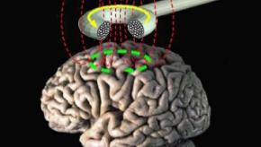
Repetitive Transcranial Magnetic Stimulation in Fibromyalgia
A randomized, sham-controlled trial has shown that repetitive transcranial magnetic stimulation (rTMS) reduced pain in fibromyalgia for up to 8 weeks; while analgesic effects waned, functional improvements remained during extended maintenance at week 16.
Read Article
ERA, APPs, & Alpha GAL (3.21.2025)
Dr. Jack Cush reviews the news and journal reports from this past week on RheumNow.com. Listen in for 2 new case questions - Ask Cush Anything.
Read Article
Increasing Prevalence of Osteoporosis
Research published in Osteoporosis International studied NHANES data showed increasing trends in osteoporosis in the United States of America. The key findings showed statistically significant results.
Read Article
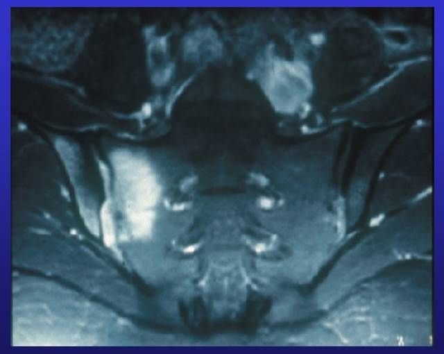
Axial Involvement in Psoriatic Arthritis
Analysis of a Greek Psoriatic Arthritis (PsA) cohort shows that nearly one quarter of patients have axial involvement, and among them, ∼30% have isolated spinal axPsA and nr-axSpA, respectively.
Increased Mortality In Arthritis Patients with COPD
People with chronic obstructive pulmonary disease (COPD) and arthritis have a higher risk of death than people with arthritis who do not have COPD, according to a new study.
Read Article
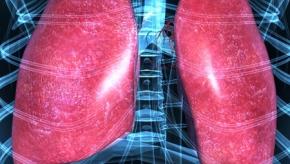
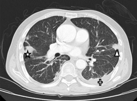
MMWR: Idiopathic Pulmonary Fibrosis Mortality in the United States
Idiopathic pulmonary fibrosis (IPF), a progressive lung disease characterized by scarring and worsening lung function, has a poor prognosis. An estimated 21% of IPF deaths might be attributable to occupational exposures.
Opioid Deaths are Down (2.28.2025)
Dr. Jack Cush reviews the news and journal reports from the past week on RheumNow.com. Opioid deaths are down, IL-33 levels are up and Weight loss is in the news again this week!
Read Article
Will AI put me out of a job?
The development of AI and other technologies brings new possibilities for optimizing diagnosis, management and has potential for improving outcomes in RA. At RheumNow Live 2025, Dr. Jeff Curtis, University of Alabama at Birmingham, presented a talk on, "AI applied to Rheumatoid Arthritis Care: Practically (Not) Perfect".
Read Article
Participation is Up! (1.31.2025)
Dr. Jack Cush reviews the news and journal reports from the past week on RheumNow.com. We also have two call-in cases on "Ask Cush Anything".
Be sure to join in the fun at RNL 2025 next weekend - Gong Show Karaoke; what will you sing?
Read Article

Eric Dein ericdeinmd ( View Tweet)

Janet Pope Janetbirdope ( View Tweet)