Understanding Non-arthritic Rheumatic iRAEs Save

A better understanding of rheumatic immune-related adverse event phenotypes beyond inflammatory arthritis has been furthered by work from three abstracts presented at EULAR 2019 in Madrid.
Ever since the emergence of immune checkpoint inhibitor immunotherapy for cancer and the rheumatic immune-related adverse events (iRAEs) they cause, questions have arisen as to what rheumatic irAEs represent, and whether they represent classical autoimmune disease or a separate entity. Increased awareness has paved the way for improved characterisation through clinical descriptions and imaging studies, which represents the basis for determining optimal therapies in the future.
Manuel Ramos-Casals, from the Hospital Clinic in Barcelona, presented work he led grouping the existing disparate reports of rheumatic irAEs published in the peer-reviewed literature into phenotypic clusters. Up until this point, the substantial majority of reports have involved case reports or small case series, which only give a limited view of phenotypes and their implications.
In Dr Ramos-Casals’s work, five phenotypic clusters were identified: articular (including inflammatory arthritis and polymyalgia rheumatica-like), muscular (including myositis and myasthenia gravis), systemic (including sicca syndrome and hemophagocytic lymphohistiocytosis), vasculitic, and granulomatous. These clusters might form the basis for more in-depth prospective characterisation, and more extensive data is likely to be available in the near future as the use of cancer immunotherapy escalates worldwide.
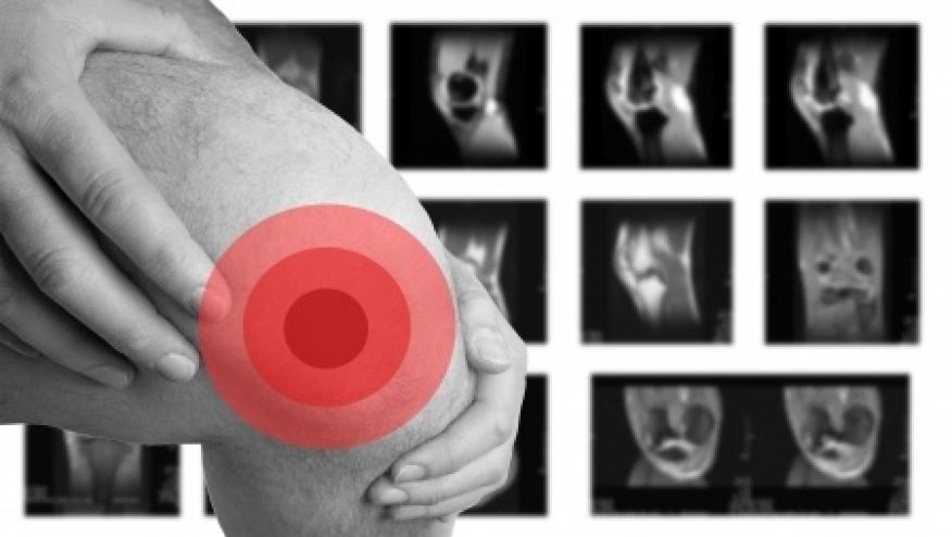
One fascinating description of a potential new irAE came from work presented by Alexandra Filippopoulou from Patras, Greece, in the form of myofasciitis noted on MRI. Periarticular manifestations have been prominent in early descriptions of rheumatic irAEs. While, of these descriptions, polymyalgia rheumatica-like disease has been most frequently described, a number of other disparate periarticular manifestations have been noted following cancer immunotherapy, although myofasciitis is not one of them.
In Dr. Filippopoulou’s study, patients with new musculoskeletal symptoms following PD-1 inhibitor immunotherapy underwent MRI scans of symptomatic areas. Most of these scans demonstrated periarticular and myofascial changes as the predominant finding, rather than synovitis, even though all these patients reported articular symptoms. This is in contrast to the predominant finding of synovitis in ultrasound studies of rheumatic irAE patients with articular symptoms, including in work also presented at EULAR 2019 by Fulvia Ceccarelli and colleagues from Sapienza Universita di Roma in Italy, and also work published online first during EULAR 2019 by Jemima Albayda and colleagues from Johns Hopkins in ACR Open Rheumatology.
It is also of interest that similar observations of myofasciitis on MRI have previously been made in classical polymyalgia rheumatica patients, although it was notable the patients described by Dr. Filippopoulou did not describe joint stiffness. These manifestations have not been noted elsewhere and further imaging studies in rheumatic irAEs elsewhere may help to delineate their importance.


If you are a health practitioner, you may Login/Register to comment.
Due to the nature of these comment forums, only health practitioners are allowed to comment at this time.