

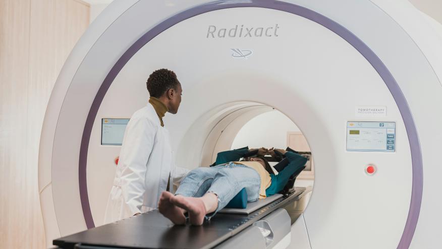
MRI as a novel biomarker in lupus Save

The opening day of #EULAR2026 brought an interesting range of topics. One can get easily overwhelmed especially for first-time attendees. Fortunately, planning early and making use of meeting resources can mitigate the confusion and ensure an enjoyable, high-yield congress attendance.
The clinical abstract sessions on diagnostic tools in lupus showcased interesting studies, including the use of MRI, as potential non-invasive biomarkers.
Abstract OP092 by Dr. C. Huang et al. aimed to evaluate the potential role of multiparametric MRI as a non-invasive tool for assessing disease activity and residual damage among patients with lupus nephritis. This prospective, single-center, observational study included patients with active (n=40) and stable lupus nephritis (n=40) and healthy controls (n=21). Qualitative imaging analysis showed two radiological phenotypes based on visible abnormalities: (1) exudative phenotype with features consistent with active inflammation and edema and (2) a non-exudative phenotype characterized by cortical thinning, scarring or low signal intensity characteristic of chronic damage. Among patients with active LN, 85% showed visible abnormalities with predominantly the exudative phenotype. In contrast, majority of patients with stable LN had non-visible abnormalities. Correlating this with renal pathology, the exudative phenotype was observed in patients with Class IV + V active LN. The quantitative analysis showed that the apparent diffusion coefficient (ADC) remained low in the exudative phenotype. Compared to healthy controls, the ADC values were also lower in stable patients suggesting irreversible interstitial fibrosis.
Meanwhile, abstract OP093 by the group of Dr. Yufei Liu aimed to characterize brain MRI findings in NPSLE patients and evaluate whether these findings have any associations with neuropsychiatric manifestations, serological profiles and clinical outcomes. Their retrospective study collected data from the Peking Union Medical College Hospital between April 2012 to January 2025. The brain MRI abnormalities were classified as inflammatory-like lesions, large-vessel diseases (LVDs), and small-vessel diseases (SVDs).
A total of 374 NPSLE patients were included in the study, majority of whom were female (85%). Abnormal brain MRI abnormalities were seen in majority of patients (66.3%) with small-vessel disease being the most prevalent (50.5%), followed by large-vessel disease (17.9%) and inflammatory-like lesions (5.9%). White matter hyperintensities and brain atrophy were the most common findings in the small-vessel diseases (SVDs). With regard to clinical associations, they found that patients with focal neuropsychiatric symptoms had abnormal MRI findings [OR 3.24 (95% CI 2.03–5.24), p < 0.001] while patients with diffuse neuropsychiatric manifestations showed normal MRIs ([OR 1.94 (1.22-3.15), p = 0.006]). In terms of serological profiles, the presence of lupus anticoagulant was associated with vascular MRI abnormalities such as white matter hyperintensities and large-vessel infarcts. Interestingly, anti-rNP antibodies had a lower likelihood of abnormal MRI findings. Prognosis-wise, the presence of abnormal MRI findings was associated with increased mortality [HR 4.5 (95% CI 1.02-19.77), p = 0.047]. In particular, brain atrophy and intracranial hemorrhage were independent predictors of mortality, ([HR 4.44 (1.89-10.45), p<0.001 and HR 4.74 (1.08-20.71), p = 0.039], respectively.
There is growing potential for innovative non-invasive imaging techniques like MRI in the evaluation and prognostication of lupus disease activity. Although limitations exist for the abovementioned studies, further research into these issues may help inform us of alternative options in monitoring or prognostic strategies in lupus.


If you are a health practitioner, you may Login/Register to comment.
Due to the nature of these comment forums, only health practitioners are allowed to comment at this time.