The Differential Diagnosis of Heel Pain Save

A slim, yoga bending, middle aged school teacher seeks a rheumatology consultation to determine the cause and cure for her left lateral heel pain for the past 6 months. No trauma, no crazy footwear and no systemic symptoms, uveitis, or peripheral/axial arthritis. Beyond inflammatory enthesopathies (Achilles tendinitis is or plantar fasciitis), the causes of heel pain are numerous. In many, the diagnostic possibilities can be established by the location and quality of the pain.
The calcaneus is the largest of 26 bones in the foot and its weight bearing role makes it a prime target for damage, entrapment or inflammation. It’s important to note that the cause of heel pain is more often mechanical than inflammatory. Heel pain has a prevalence of 3.6% and is said to affect over 2 million Americans. The following may be considered in the evaluation of heel pain.
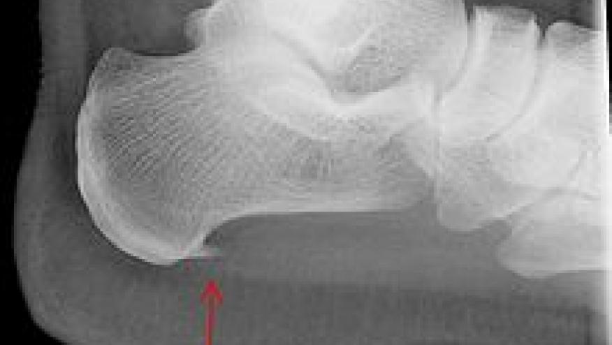
Calcaneal Fractures: Stress fractures usually manifest as progressive pain following an increase in activity level or walking on hard surfaces. These are often due to repetitive stress, strenuous exercise/sports (especially runners) or heavy manual work. At risk individuals include runners (may also get metatarsal stress fracture) and those with osteoporosis. These are diagnosed by MRI (early) or plain radiography (late) and usually located inferior and posterior to the posterior facet of the subtalar joint. Clues include pain at rest, local ecchymosis, point tenderness at the fracture site.

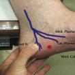
Entrapment Neuropathy: Nerve entrapment can be suggested by pain or associated neuropathic symptoms - burning, tingling, or numbness. Such neuropathies are often unilateral, involving branches of posterior tibial nerve - the medial plantar nerve, lateral plantar nerve (also known as the nerve to the abductor digiti minimi) or the lateral calcaneal nerve (off the sural nerve). Lumbar radiculopathies involving L4-S2 may also underlie neuropathic heel pain.
Haglund Deformity: Posterior heel pain can also be attributed to a Haglund deformity, a bony prominence over the superior posterior calcaneus that may lead to bursitis between the calcaneus and Achilles tendon. It is most commonly seen in women in their twenties. Heel Pad Syndrome: This syndrome is common in older individuals and is felt as a deep, bruise-like pain in the central heel and can be induced by manual pressure or walking barefoot on hard surfaces. It is often associated with fat pad atrophy.
Tarsal Tunnel Syndrome: The tarsal tunnel is formed by the flexor retinaculum, medial calcaneus, posterior talus, and medial malleolus. Compression of the posterior tibial nerve as it courses through the tunnel leads to neuropathic pain and numbness in the posteromedial ankle/heel or medial midfoot. The diagnosis can be established by electrodiagnostic testing or Tinels sign (thumping over the posterior tibial nerve).
Sinus Tarsi Syndrome: Pain arise from the sinus tarsi that includes the space between the calcaneus, talus, and talocalcaneonavicular and subtalar joints. The pain is felt over the lateral calcaneus/heel or lateral midfoot. Causes include excessive motion of the subtalar joint, hyperpronation or subtalar instability.
This particular patient had lateral calcaneal pain. Based on the above, her differential diagnosis may include calcaneal stress fracture, entrapment neuropathy (lateral plantar nerve) or the sinus tarsi syndrome.


If you are a health practitioner, you may Login/Register to comment.
Due to the nature of these comment forums, only health practitioners are allowed to comment at this time.