


Evaluating Inflammatory Joint Pain in Older Adults Save

Know-it-now
- Inflammatory arthritis (IA) diagnostic delays are longer (1 year) in older adults due to atypical presentations or symptoms ascribed to aging
- Seronegative RA mimicking PMR is common in late-onset disease
- True synovitis (hands/wrists), elevated inflammatory markers, and progressive dysfunction favors RA over PMR
- RS3PE and atypical/refractory IA warrant occult malignancy screening
- Imaging: radiography can be supplemented with ultrasound or MRI, if IA is suspected but clinically equivocal
- Age is not a contraindication to DMARD therapy and glucocorticoids have serious risks and should be minimized and rapidly tapered
Lee, Levinson, and Makris have published an approach to evaluating inflammatory joint pain in older adults in JAMA Internal Medicine. While intended to be a practical guide for Primary Care Clinicians (PCCs), the tenets and clues ring true for all.
Arthritis affects nearly one-third of US adults ≥65 years, and while osteoarthritis (OA) predominates, inflammatory arthritis (IA)—rheumatoid arthritis (RA), spondyloarthritis, crystal arthropathies, and related conditions—is often missed, largely because of atypical, overlapping presentations, or symptoms attributed to aging.
IA features morning stiffness >1 hour, nocturnal pain, and improvement with activity, versus OA's worsening with use and relief with rest. Constitutional symptoms (weight loss, fatigue, poor appetite) should not be attributed to normal aging. (Editors note: IA liklihood goes up with chronicity [>12 weeks of joint pain/swelling], the four cardinal signs of inflammation and lab evidence [ESR or CRP or anemia or thrombocytosis] of inflammation).
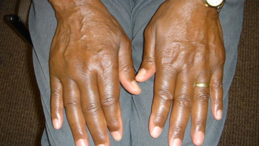
Mimics. RA can present de novo in the seventh–eighth decade, is often seronegative and shoulder/large-joint predominant—mimicking polymyalgia rheumatica (PMR), which itself causes no true synovitis. Crystal arthropathies present as acute/subacute mono- or oligoarticular flares (gout favoring podagra, knees, wrists; CPPD favoring knees/wrists and sometimes mimicking polyarticular RA). New-onset spondyloarthritis after age 70 is uncommon. RS3PE is a rare disorder, but presents as bilateral hand synovitis with dorsal pitting edema, typically seronegative and steroid-responsive—warrants a paraneoplastic workup.
Diagnostics. IA is a clinical, symptom based diagnosis that can be fortified or mislead by investigations. ESR may be physiologically elevated (normally 30-40 mm/hr in those over 70yrs.) in healthy older adults or those with anemia, so interpret in context. RF/anti-CCP are seronegative more often in this population; ANA and HLA-B27 should be reserved for suggestive phenotypes, as isolated positives are rarely diagnostic. Imaging (ultrasound or MRI) is favored over plain radiography when IA is clinically uncertain, since radiographic OA commonly coexists. Any acute monoarticular effusion warrants prompt aspiration and assessments for infection or crystal induced arthritis. In the elderly, septic arthritis may lack fever or leukocytosis.
Treatment guidance. For some unfortunate bias, elderly patients with IA may not receive warranted disease modifying antirheumatic drug (DMARD) therapy. DMARD initiation should not be delayed by age when IA is strongly suspected and rheumatology access is limited. Methotrexate remains first-line at 10-20 mg weekly with folic acid; dose-adjust below eGFR 60 mL/min/1.73m², avoid below 30. Hydroxychloroquine is capped at ≤5 mg/kg/day (max 400 mg) with annual ophthalmologic monitoring. Glucocorticoids—even <5 mg/day prednisone—carry elevated risks of infection, osteoporosis, diabetes, hypertension, delirium, and impaired wound healing; taper 25%-50% weekly toward bridging use, targeting ≤5 mg/day if ongoing use is unavoidable, with bone/vaccination/metabolic monitoring beyond 3 months. Biologics and targeted synthetic agents (JAK inhibitors) are equally effective and safe in young and older IA patients.
This article is a useful referral-pattern primer—reinforcing that seronegative, large-joint RA and PMR-mimicking presentations deserve a low threshold for synovitis confirmation (exam, ultrasound, or MRI) before anchoring on PMR, and that occult malignancy screening belongs in the workup of atypical or refractory IA patients.
Related Content


ADD THE FIRST COMMENT
Disclosures
Disclosures
The author has no conflicts of interest to disclose related to this subject
The author used AI to research and organize this content, and maintains responsibility for its accuracy




If you are a health practitioner, you may Login/Register to comment.
Due to the nature of these comment forums, only health practitioners are allowed to comment at this time.