

Top 5 and Bottom 5 Joints (Best of 2015) Save

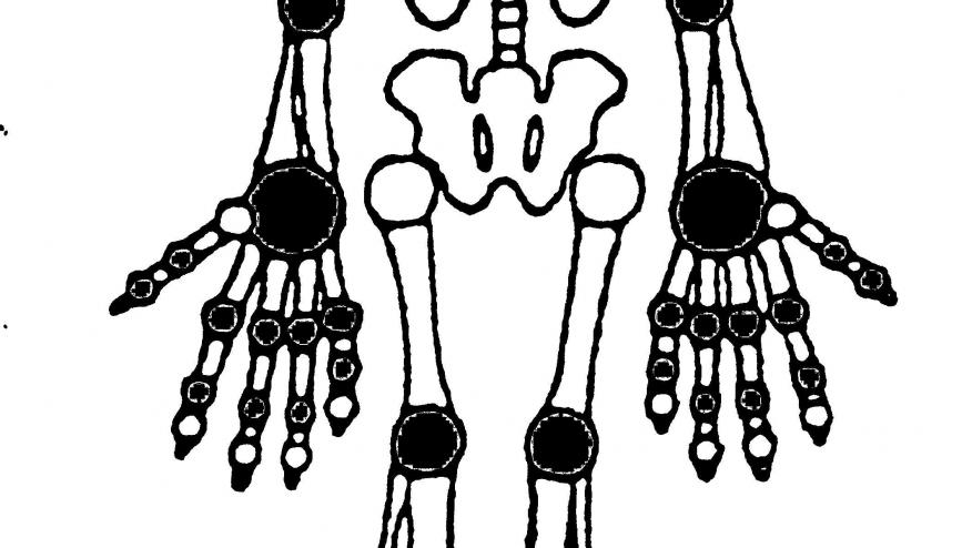
Who knows or ponders the articulations (joints) more so than the rheumatologist or orthopedist? We know their anatomy, their contours, tender sites and how to examine them. Adults have over 200 bones and up to 360 joints. Imagine if you had to do a DAS360?
They say if you are going to spend one-third of your life sleeping, you should probably invest in a good bed to be good at sleep. In the same vein, if you’re going to spend 10-20% of your clinic hours examining and caring for 28 or 68 or 360 joints – it may be instructive to own up to those joints we excel at and take note of the ones we avoid, despise or struggle with. Now, don’t play proud parent and declare you love them equally. No one believes you, including your cricoarytenoid joint.(The following listing represents the views of the author and does not represent the views of the RheumNow staff, faculty or other rheumatologists round the world.)
My Bottom 5 Joints
5. Zygapophyseal: Seldom mentioned and then only in dimly lit radiology reading rooms, the zygapophyseal joint is inaccessible, unspellable (hence the term Z-joint), and has a changing orientation up and down the spine – a miserably inconsistent joint. And that’s only half the problem. These facet joints play a significant role in society’s #1 public health ache – back pain. There’s a whole economy based on injection of the Z-joint (by ortho and pain specialists), yet evidence-based studies show these are less effective than chiropractic manipulations. Wait…one more, chiropractors love to point to these unmentionable joints on X-rays, while they explain their approach to the ailing patient, who wants relief more so than a lecture on roentgenographic anatomy gone wrong.
4. Hip: I’m too am surprised to put the hip joint in this pile. So I understand your “what-tha” objections. But my reasons are sound, even if my evidence is scant. First, patients and doctors frequently talk about the “hip” joint or pain and we rarely are discussing the same origin or articulation. In my experience, of the patients who talk of hip pain, 80% are plagued by pain over the gluteal region (referred from lumbosacral pathology), 15% point to their lateral trochanteric pain (caused by either bursitis or fibromyalgia) and only 5% point to deep anterior groin/inguinal pain (from the true hip joints). To properly assess the hip, the patient must lie on the exam table. Come on man! A rheum patient on an exam table is usually not a good thing. Patients may be sicker, or at least believe they are. An uncomfortable exam or procedure soon follows. I may have to wear gloves or perform the FABERE-4 exam or some other poorly predictive maneuver. Granted, there’s much good that can come from a rheumatologist and the hip exam, but in my mind it belongs on my bottom five joint.
3. Shoulder: This large joint is the cause of large problems. Degenerative, traumatic, occasionally inflammatory and rarely infectious, the shoulder has numerous etiologies, and it has numerous origins: the glenohumeral, acromiclavicular, bicipital tendonitis, impingement syndrome, rotator cuff disturbances, referred cervical pain, fibomyalgia/myofascial pain, polymyalgia rheumatica, etc. If I had an ultrasound and knew how to use it, I would probably love the shoulder joint – but I’m $30,000 short and 30 years too old to turn this one into a top five joint.
2. Temporomandibular: Easy: a) most with painful TMJs have fibromyalgia, myofascial pain or poor sleep; b) TMJ pathology may be tied to dental strife and mastication problems; and C) the TMJ clinical exam requires you to put your index finger deep into the ear (a surprise feeling for both patient and MD) and your thumb on the base of the mandible while the patient opens and closes the mouth. This is an odd exam that is largely unrevealing and overly moist.
1. Sacrococcygeal symphysis: Hah! You didn’t even think of this one and didn’t know this amphiarthrosis articulates the sacrum and coccyx. Other than a complicit role during childbirth, I don’t know what purpose it serves. If a patient points down there, I’m afraid to follow that lead. If I had to examine it, would it be like the TMJ exam? Would I have to wear gloves to get the full perspective? All kinds of crazy things go on down there – teratomas, fistulas, cysts! This one doesn’t come up much, thankfully.
My Top 5 Joints
5. Sacroiliac: Rheumatologists are enamored with the SI joint. Although hardly accessible and not easily examined, the finding of an abnormal SI arthritis takes on numerous potential etiologies that excites either a division of rheumatologists or partners in practice to ponder the benefits or cure that will ensue. Low back pain is just that. However, inflammatory low back pain with an abnormal SI X-ray ensures far better outcome than that seen with other causes of back pain.
4. Knee: Our classic diarthrodial hinge joint, where good clinical skills and experience alone can diagnose osteoarthritis, inflammatory arthritis, crystal or septic arthritis, meniscal or cruciate tears, etc. This is our best see-one, do-one, teach-one joint. The magic of arthrocentesis, ultrasound and radiology seems optimal with the knee more so than any other. And just when things get really bad, we can scope it or partially or fully replace the joint and give our patients a whole new lease on life!
3. Wrist: This one is about as high tech as any part of the body. Sixteen bones converging beneath your wrist watch, with innumerable articulations that are strong enough to protect yourself and nimble enough to perform a drum solo or hang a curve ball. All the while serving as a conduit for the plumbing and electric supply to the hand and fingers.
2. Distal interphalangeal: Easy to inspect and diagnose is the DIP. Most of what happens here is instantaneous. It’s often the first joint we squeeze. If this one hurts it’s either going to be osteoarthritis or a long joint exam. This also happens to be the joint whose visual appearance troubles patients most. Such a small joint can have large consequences and lead to impactful discussions with the patient. Unfortunately, limited treatment options keep it out of the top slot.
1. Metacarpophalangeal: It doesn’t get any better than the MCP; a joint that defines the skill and compassion of the rheumatologist and encompasses why rheumatologists are the happiest of specialists. Face to face with the patient, the rheumatologist gently holds the hand, asking and discerning the patient’s health. With two hands, he/she holds the patient’s hand, and with four finger tips circumferentially slide and sense (with MRI like precision) the contours, content, stability and state of that one joint, all the while watching the patient’s face for a wince or reaction. Most medical specialties don’t get to spend much time hand holding. There is something truly caring about the hand exam. Rheumatologists should slow down at the hands to appreciate what they are doing and to show how much they care. For those who’ve done thousands of exams and can’t appreciate slight synovial swelling or the patient connection, I can only recommend retirement or becoming an endocrinologist where palpation and inspection of lab reports (thyroid function tests, HgbA1c) is as good as it gets.
ADD THE FIRST COMMENT
Disclosures
The author has no conflicts of interest to disclose related to this subject


If you are a health practitioner, you may Login/Register to comment.
Due to the nature of these comment forums, only health practitioners are allowed to comment at this time.