

COMMON - Incidental Rotator Cuff Abnormalities on MRI Save

In this cross-sectional study of 602 Finnish adults undergoing bilateral shoulder magnetic resonance imaging (MRI) and clinical assessment, found abnormalities in nearly everyone over age 40yrs, regardless if asymptomatic or symptomatic.
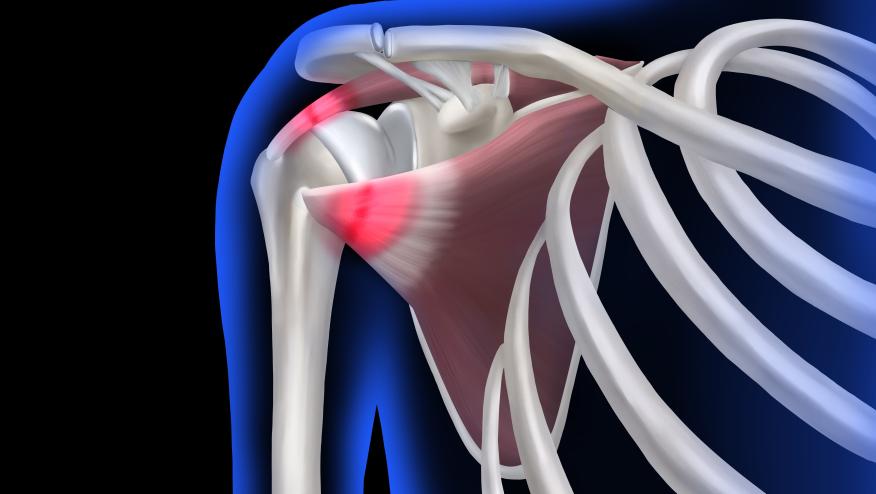
Shoulder pain leads to a consideration of rotator cuff (RC) abnormalities. Should diagnostic imaging be used, including MRI?
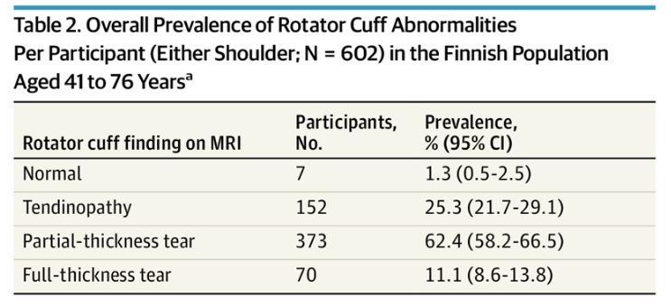
A population-based cross-sectional study evaluated adults aged 41 to 76 years who underwent standardized clinical assessment and bilateral 3-Tesla magnetic resonance imaging (MRI) of the shoulders. RC tendon status was classified on MRI as normal, tendinopathic, partial-thickness tear (PTT), or full-thickness tear (FTT).
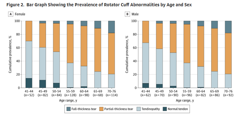
Among 602 participants (median age, 58 [range, 41-76] years; 52% female), MRI RC abnormalities were found in 595 (98.7%; 95% CI, 97.5%-99.5%): 25% tendinopathy, 62% PTT, and 11% FTT. The prevalence and severity of abnormalities increased with age, but did not differ between sexes.
RC abnormalities were present in 96% of asymptomatic and 98% of symptomatic shoulders. Only FTTs were more prevalent in symptomatic shoulders (14.6% vs 6.5% in asymptomatic shoulders).
The findings of this study suggest that RC abnormalities are nearly universal after age 40 years and that routine imaging should not guide diagnosis or treatment of atraumatic shoulder pain.
Continue Reading
ADD THE FIRST COMMENT
Disclosures
The author has no conflicts of interest to disclose related to this subject


If you are a health practitioner, you may Login/Register to comment.
Due to the nature of these comment forums, only health practitioners are allowed to comment at this time.