

Lupus Hepatitis and Lupoid Hepatitis Reviewed Save

SLE-related hepatitis (“lupus hepatitis”) and “lupoid hepatitis” are 2 different diseases. The term "lupoid hepatitis", now known as autoimmune hepatitis (AIH), was first coined in 1959. AIH was initially called “lupoid hepatitis” by MacKay. A review by Adiga and Nugent reviews, defines and distinguishes between lupus-related hepatitis and lupoid hepatitis.
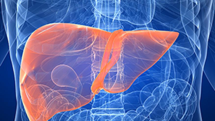
Liver dysfunction occurs in up to 50% of patients with systemic lupus erythematosus (SLE) and transaminitis is seen in over a quarter patients. Such findings pose a diagnostic challenge in many SLE patients as elevated liver enzymes (LFTs) have a wide differential diagnosis; ranging from hepatitis (infectious, lupoid, autoimmune, toxin/drug-induced) to other forms of hepatocellular or cholestatic disease and muscle damage.
Distinguishing between lupus hepatitis and lupoid hepatitis (autoimmune hepatitis [AIH]) can be a challenge as both primarily affect women and share other features like, hypergammaglobulinemia, autoantibody (ANA) positivity and a response to immunosuppressive therapy.
While the majority of patients with liver disease will have autoantibody positivity, this does not equate to an SLE diagnosis. Moreover, nearly 25% of AIH patients will fulfill criteria for SLE.
Serology may help distinguish between the lupus and AIH; wherein anti-SLA, anti-SMA and anti-LKM antibodies are found in AIH and low complement, ribosomal P antibodies can been seen in SLE (Table).
Liver biopsies are often necessary to establish the diagnosis of lupus hepatitis and AIH. Lupus hepatitis has lobular infiltrates, mild portal infiltration with lymphocytes, neutrophils and plasma cells, steatosis, complement 1q deposition. mild cholestasis, focal necrosis and nodular cirrhosis can also be seen. AIH demonstrates portal infiltrates with periportal piecemeal necrosis, hepatocyte rosettes, bridging fibrosis, panlobular/multilobular necrosis and cirrhosis suggest AIH.
| Lupus hepatitis | Autoimmune hepatitis | |
|---|---|---|
| ANA positivity | Most of the patients | 80% in type 1 autoimmune hepatitis |
| Complement | Low | Normal |
| Anti–SMA antibody | May be positive | Usually positive in type 1 autoimmune hepatitis |
| Anti–LKM antibody | Negative | Positive in type 2 autoimmune hepatitis |
| Anti–liver-pancreas antigen | Negative | Positive in type 3 autoimmune hepatitis |
| Anti–ribosomal P antibody | Positive | Negative |
| Histology | Lobular infiltrates with paucity of lymphocytes. Mild chronic inflammation | Periportal piecemeal necrosis and hepatocyte rosette formation. Abundant plasma cell and lymphocyte infiltrate |
| Progression | Benign | Progresses to cirrhosis |
| Prognosis | Good | 5-Year survival 80% in treated patients and 25% in untreated patients |
*AMA, antimitochondrial antibody; ANA, antinuclear antibody; anti-LKM, antibody to liver or kidney microsomes; anti-SMA, anti–smooth muscle antibody.
Lupus Hepatitis. Transaminitis occurs in 25-59% of lupus patients. Such LFT elevations may be drug-induced (ASA, azathioprine, methotrexate antimalarial drugs), or due to nonalcoholic fatty liver disease or nonalcoholic steatohepatitis, or viral hepatitis. With these excluded, 28-42% had lupus hepatitis.
Autoimmune Hepatitis. AIH is a chronic hepatitis of unknown etiology characterized by hepatocellular necrosis and inflammation; it affects 100,000-200,000 persons in the United States. AIH may present as abnormal LFTs or can present with acute, sometimes fulminant hepatitis, with associated nausea, anorexia, abdominal pain and jaundice, with or without other lupus symptoms or other autoimmune phenomenon (ITP, autoimmune thyroiditis, synovitis, ulcerative colitis).
Type I AIH is the classic syndrome seen in young women and is associated with marked hypergammaglobulinemia, lupoid features and positive antinuclear antibodies (ANAs). Type II AIH is usually seen in children and in Mediterranean populations and is associated with anti–liver and kidney microsomal antibodies and a negative ANA. Type III AIH is associated with positive ANA, smooth muscle antibody (SMA) and antibodies to soluble liver antigen or liver-pancreas (anti-SLA/LP).
AIH has a more aggressive histology compared to lupus hepatitis, and untreated symptomatic AIH has a poor prognosis with a 5-year survival rate less than 25% in untreated patients versus 80% in those treated with corticosteroids.
Continue Reading
ADD THE FIRST COMMENT
Disclosures
The author has no conflicts of interest to disclose related to this subject


If you are a health practitioner, you may Login/Register to comment.
Due to the nature of these comment forums, only health practitioners are allowed to comment at this time.