

Spinal Findings on MRI May Not Signify Spondyloarthritis Save

For about one in six people considered healthy with no reports of back pain, lesions were visible on MRI in the lower spinal column and sacroiliac joint, researchers said -- a sign that rheumatologists should be cautious about diagnosing spondyloarthritis (SpA) on the basis of imaging.
Out of 95 participants in the study, SpA-type lesions were seen in 17, according to Thomas Renson, MD, PhD, of Ghent University Hospital in Belgium, and colleagues. Yet only one had multiple corner inflammatory lesions indicative of an autoimmune process, and none of the 17 were positive for HLA-B27, an SpA biomarker in blood.
"Contrary to what is commonly believed, structural MRI-detected SI joint lesions are frequently seen in healthy individuals," the researchers reported in Arthritis & Rheumatology. Most of the MRI-detected lesions were in individuals 40 and older, the group noted.
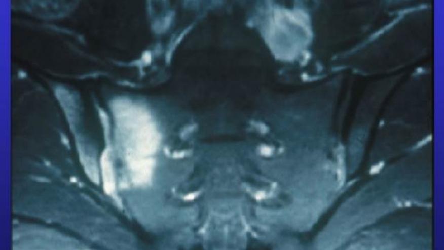
MRI is the "gold standard" for detecting inflammation of the sacroiliac joint, which in turn is a "hallmark" of axial SpA, Renson and colleagues observed. Yet a number of other research groups have reported substantial prevalence of imaging abnormalities in people unlikely to be suffering from SpA, such as young women, military recruits, and athletes. How common such findings might be in the general population isn't known. Thus, the Ghent group organized what they said was the first-ever study to examine the issue specifically in people without back pain.
Renson and colleagues recruited 95 adults (in roughly equal numbers for ages 20-29, 30-39, and 40-49) from among Ghent University Hospital staff and their own relatives and acquaintances, all of whom said they had no current back trouble. Three indicated they had previously experienced chronic back pain but it had eased.
In addition to MRI directed to the pelvic region, participants provided blood samples and completed a brief questionnaire about their overall health, medical history for themselves and family, and medication use. Those using nonsteroidal anti-inflammatory drugs in the previous 2 weeks or having had treatment with a tumor necrosis factor inhibitor were excluded.
MRI was performed on a 3T machine and images were analyzed for the presence of deep bone marrow edema (BME), fat metaplasia, erosion, and new bone formation. The researchers also applied the Spondyloarthritis Research Consortium of Canada (SPARCC) scoring system, and determined whether the findings met Assessment of SpondyloArthritis International Society (ASAS) criteria for SpA positivity.
One-fifth of participants showed sacroiliac joint erosions, and 14% had fat metaplasia. Erosions were seen in nearly 40% of those 40 and older; spinal BME was also present in 36% of this age group, and 29% showed fat metaplasia. SPARCC scores for both the sacroiliac and spine correlated with age. Among the subset 45 and older, the 95% confidence intervals for SPARCC scores overlapped those for a historical cohort of 84 SpA patients.
But only one participant had at least three corner inflammatory lesions, which would be more highly indicative of SpA. Meanwhile, among the three with previous back pain episodes, all had SPARCC scores of 0. And none of the participants with MRI-detected lesions were positive for the HLA-B27 biomarker.
Renson and colleagues said the findings of joint erosions and fat metaplasia even in participants younger than 40 "challeng[es] the interpretation of [sacroiliac] joint MRI in patients with a clinical suspicion of SpA."
Furthermore, they asserted, the study results stand "in contrast with the general belief that structural MRI-detected [sacroiliac] joint lesions are specific for SpA and can contribute to a diagnosis of [axial] SpA, particularly in patients with suggestive symptoms but without BME" on MRI.
The investigators also found that participants with erosions tended to have higher body mass index values. They noted, too, that those with overweight and obesity typically show higher levels of C-reactive protein relative to those of normal weight. This, Renson and colleagues wrote, "further complicat[es] the distinction of SpA in overweight individuals with chronic back pain."
Limitations to the study included the small number of participants, making subgroup comparisons inconclusive.
Continue Reading
ADD THE FIRST COMMENT
Disclosures
The author has no conflicts of interest to disclose related to this subject


If you are a health practitioner, you may Login/Register to comment.
Due to the nature of these comment forums, only health practitioners are allowed to comment at this time.