

2023 EULAR Recommendations on Imaging in Crystal-Induced Arthritis Save

New recommendations on imaging for crystal-induced (gout, CPPD) arthritis (CiA) were presented at EULAR 2023 in Milan. These included 5 overarching principles & 10 recommendations on the role of imaging in making a diagnosis, monitoring, predicting, guiding therapy, and patient education in CiA.
The task force included 25 stakeholders from 11 countries, who performed four systematic literature searches answering 14 research questions on the role of imaging in gout, calcium pyrophosphate and basic calcium phosphate deposition disease. Key symptomatic and imaging joints include the first metatarsophalangeal joint in gout, the wrist and knee in CPPD, and the shoulder in BCPD.
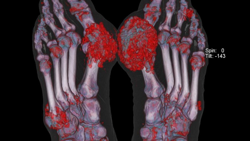
Notably, if classic findings of gout are found on imaging, synovial fluid analysis is not necessary to confirm the diagnosis. Also, the ultrasound and dual-energy CT scanning are discussed.
Overarching Principles
- CiA are typically characterized by intermittent, acute episodes of inflammation, but may also exhibit a persistent or indolent disease with or without superimposed flares
- Imaging in CiA provides useful information on crystal deposition, inflammation, and structural damage
- Imaging abnormalities may not always be related to clinical manifestations in CiA
- Patient information (medical history, physical/laboratory examination, synovial fluid/tissue analysis) should be taken into account when imaging is considered in CiA
- Imaging in CiA should be performed and interpreted by trained health care professionals.
Recommendations
- When performing imaging in CiA, both symptomatic areas and disease-specific target sites (i.e. MTP1 in gout, wrist and knee in CPPD, shoulder in BCPD) should be considered
- In the diagnostic assessment of gout, US and DECT are both recommended imaging modalities
- When characteristic features of MSU crystal deposition on US (i.e. double contour sign or tophi) or on DECT are identified, synovial fluid analysis is not needed to confirm a diagnosis of gout
- In the diagnostic assessment of CPPD, CR and US (or CT if axial involvement is suspected) are recommended imaging modalities
- In the diagnostic assessment of BCPD, imaging is necessary; CR or US are the recommended modalities
- In gout, US and DECT can be used to monitor crystal deposition and in case of US, also inflammation. Both modalities provide additional information on top of clinical and biochemical assessment. In case US/DECT are not available, CR can be used to assess structural damage due to gout. The decision on when to repeat imaging depends on the clinical circumstances
- In CPPD and BCPD serial imaging is not recommended, unless there is an unexpected change in clinical characteristics
- In gout, assessing the amount of MSU crystal deposition by US or DECT may be used to predict future flares
- If synovial fluid analysis is required in the assessment of CiA, US-guidance should be used in cases where aspiration based on anatomical landmarks is challenging
- Showing and explaining imaging findings of CiA to people with such conditions may help them understand their condition and improve treatment adherence in gout.
Join The Discussion
The availability of DECT is very limited in the UK.
CR: conventional radiography
like
Disclosures
The author has no conflicts of interest to disclose related to this subject


If you are a health practitioner, you may Login/Register to comment.
Due to the nature of these comment forums, only health practitioners are allowed to comment at this time.