

Predictive FDG-PET in Newly Diagnosed GCA Save

Annals of Internal Medicine reports that the use of 18F-fluorodeoxyglucose (FDG) PET imaging at the time of a giant cell arteritis (GCA) diagnosis may help estimate the risk for aortic aneurysm formation.
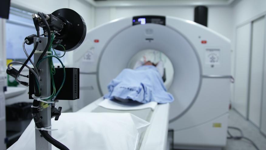
A pospective cohort study recruited 106 GCA patients from the University Hospitals Leuven. GCA patients underwent FDG positron emission tomography (PET) imaging within 3 days of glucocorticoid initiation and thereafter computed tomography (CT) imaging yearly for a maximum of 10 years. PET scans were scored (0 to 3) in 7 vascular areas and summed to a total vascular score (TVS).
Overall, patients with a positive PET scan result at diagnosis had greater increases in ascending aorta diameter at 5-years. Thoracic aortic dimensions were also positively associated with TVS at onset.
The risk of thoracic aortic aneurysms was significanly increased in those with a positive PET scan result (adjusted hazard ratio, 10.21 [CI, 1.25 to 83.3]).
Higher TVS was associated with greater yearly increase in thoracic aortic dimensions. Performing PET imaging at diagnosis may help to estimate the risk for aortic aneurysm formation.
ADD THE FIRST COMMENT
Disclosures
The author has no conflicts of interest to disclose related to this subject


If you are a health practitioner, you may Login/Register to comment.
Due to the nature of these comment forums, only health practitioners are allowed to comment at this time.