Using MRI to Evaluate Knee Pain Save
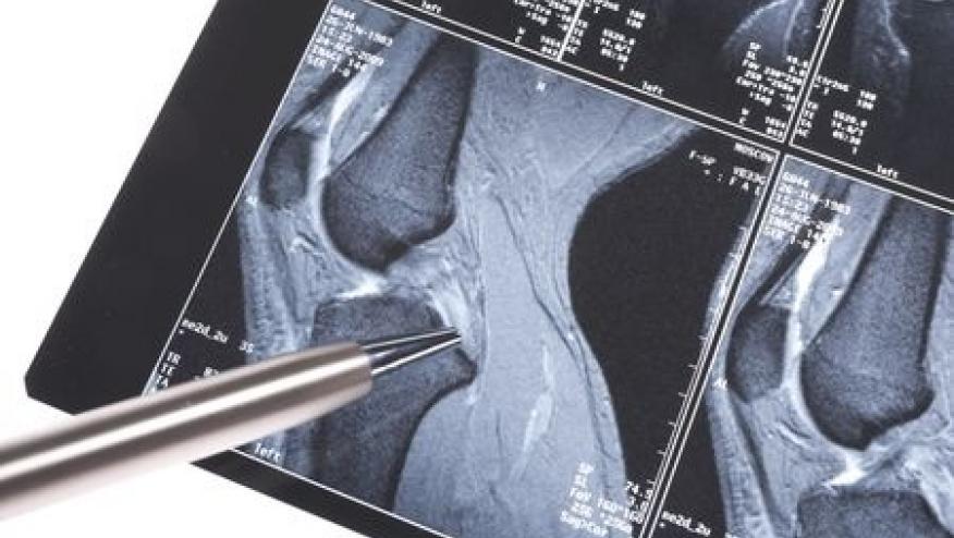
Interim results from a randomized trial show that middle-aged patients with knee pain and suspected meniscal tears can benefit from MRI scans even in relatively simple cases.
MRI findings from 760 such patients showed that 61 (8%) had issues other than plain meniscal tears that, in normal clinical practice, would alter their management, according to Stacy E. Smith, MD, of Brigham and Women's Hospital in Boston, and colleagues in Arthritis Care & Research.
Among these were 25 cases of subchondral insufficiency fracture of the knee (SIFK) -- a condition that is normally treated with rest, whereas physical therapy involving leg exercise is commonly prescribed for meniscal tears.
Other disorders identified from the scans included tumors, avascular necrosis, and a variety of even less common conditions such as bipartite patella and intraosseous ganglion.
These findings, Smith and colleagues suggested, "may prompt clinicians to be more aware" of these possibilities that can only be discovered with advanced imaging. "Future research is needed to pinpoint factors associated with these concerning findings, so that patients at highest risk can be identified and referred for advanced imaging without delay."
Normally, patients with osteoarthritis of the knee and suspected meniscal tears are diagnosed from clinical signs and x-rays. MRI scans, Smith's group noted, are typically reserved only for "complex cases" in light of the vastly greater cost. But as the study ultimately confirmed, a minority of such patients will have other causes for their symptoms for which MRI scans are better suited to detect. The question is, how big (or small) of a minority?
A unique data source to provide an answer is the ongoing TeMPO study (Treatment of Meniscal Tears and Osteoarthritis), a treatment trial that began in 2018 testing four approaches to these common problems. (The trial was completed last October with results yet to be reported.) Enrollment procedures included an MRI scan at baseline to confirm the diagnosis of meniscal tear. Thus, Smith and colleagues had a population of relatively ordinary patients with suspected tears who would all undergo MRI scans, and thus could provide an estimate of the prevalence of alternate pathologies.
Mean patient age was about 60, and some 40% were men. BMI values averaged about 30. Patients scored their degree of knee pain at an average of roughly 50 on a 100-point scale.
MRI findings showed that 18% of the sample had no meniscal tear, but in about half of that group the scans didn't reveal anything else of clinical importance. Meanwhile, among the 61 with "concerning" alternative findings, meniscal tears were confirmed in more than half.
Besides the 25 patients with SIFK, 10 had other types of fractures. Additionally, the imaging detected tumors and osseous lesions in eight patients, avascular necrosis in four, and 14 with "other" significant conditions. Most common in the "other" category was bone contusion or edema lacking a fracture line, seen in seven cases; osteochondral lesions were seen in three patients.
Importantly, Smith and colleagues did not recommend that all patients with suspected meniscal tears undergo MRI scans. Rather, advanced imaging should be reserved for those at increased risk for having other explanations for their symptoms. However, because relatively few patients in the current study had these alternative diagnoses, the researchers couldn't make much headway in identifying useable risk factors.
Other limitations included the study's conduct at academic centers in Boston, Buffalo, and Pittsburgh, perhaps reducing the generalizability to other regions with different patient populations.
Source Reference: Waddell LM, et al "Prevalence of clinically relevant findings on MRI in middle-aged adults with knee pain and suspected meniscal tear: a follow-up" Arthritis Care Res 2024; DOI: 10.1002/acr.25444.




If you are a health practitioner, you may Login/Register to comment.
Due to the nature of these comment forums, only health practitioners are allowed to comment at this time.