

Diagnostic Yield of Cranial and Large Vessel Imaging in GCA Save

Combined cranial and large vessel ultrasound and PET/CT provided excellent accuracy for the diagnosis of giant cell arteritis (GCA).
Current guidelines call for imaging of extracranial vessels with the diagnosis of GCA - but with which imaging modality?
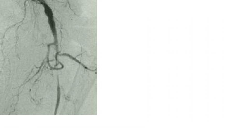
A metanalysis of 14 studies and 1727 patients examined the diagnostic accuracy of ultrasound, PET/CT and MRI. Findings included:
- Ultrasound: combined cranial and large vessel ultrasound had a sensitivity of 86% (76-92%) and specificity of 96% (92-98%).
- PET/CT of both cranial and large vessels yielded a sensitivity of 82% (61-93%) and specificity of 79% (60-90%).
- No studies assessed both PET/CT and ultrasound (thus, no head-to-head comparisons)
- Adding large vessel ultrasound to temporal artery ultrasound significantly increased sensitivity (91% versus 80%, p < 0.001) without decreasing specificity (96% versus 95%, p = 0.57).
- Evaluating cranial arteries in addition to large vessels on PET/CT increased the sensitivity (82% versus 68%, p = 0.07) without decrease in specificity (81% versus 79%, p = 0.70)
- There was insufficient data on MRI
Either PET/CT or ultrasound of extracranial vessels may be included in the evaluation of GCA patients, depending on setting, expertise, clinical presentation and imaging availability.
ADD THE FIRST COMMENT
Disclosures
The author has no conflicts of interest to disclose related to this subject


If you are a health practitioner, you may Login/Register to comment.
Due to the nature of these comment forums, only health practitioners are allowed to comment at this time.