

JAMA: Review of Shoulder Pain Save


Know-it-now
- Adopt "subacromial pain" over structural labels — pathoanatomic diagnoses overstate certainty that imaging cannot support.
- Reserve imaging for red flags (infection, malignancy, trauma, progressive neurologic deficit, worsening course) — not routine workup, especially in patients over 60, where incidental findings are the rule rather than the exception.
- Special orthopedic tests are unreliable — base diagnosis on history, active/passive range of motion, and lag signs, not isolated Neer/Hawkins/Speed results.
- Surgery has a limited, well-defined role — high-certainty evidence excludes benefit for subacromial decompression, and rotator cuff repair benefit remains uncertain outside specific indications.
- First-line care is uniform across most shoulder diagnoses — education, self-management, and watchful waiting, with glucocorticoid injection reserved for inadequately controlled pain.
JAMA Internal Medicine (Haas et al, 2026) has published a clinical overview to the approach, diagnosis and management of Shoulder Pain.
Shoulder pain affects 16% of adults annually and is the third most common musculoskeletal presentation in primary care. This new review reframes the diagnostic and therapeutic approach, moving away from pathoanatomic labels (rotator cuff tendinopathy, impingement, bursitis) toward the umbrella term subacromial pain, since structural findings on imaging correlate poorly with symptoms and are common in asymptomatic people. Roughly 90%–95% of primary care shoulder pain is subacromial in origin.
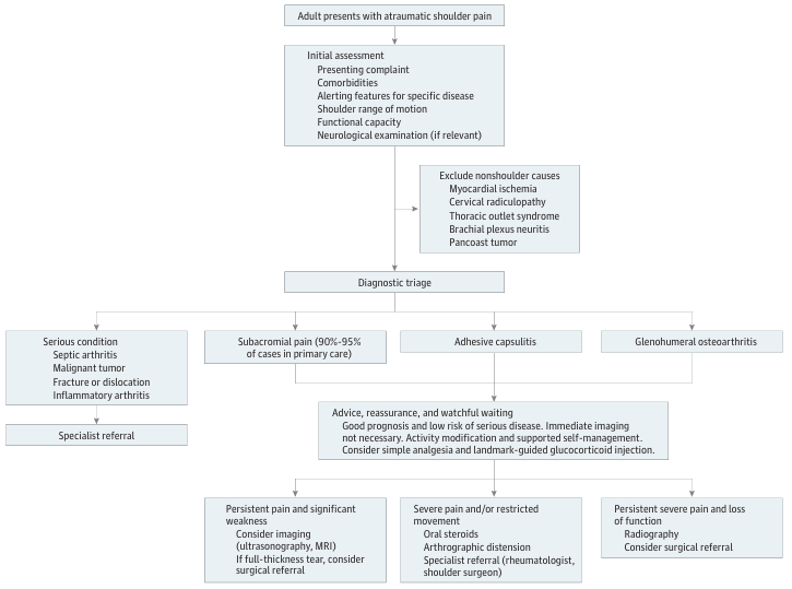
Diagnosis. Distinguishing true shoulder pathology from referred pain is a critical first step. It's important to know the anatomic origin of patients shoulder pain to know the differential diagnoses:
Shoulder: true shoulder conditions include subacromial pain, biceps tendinopathy/rupture, calcific tendinitis, adhesive capsulitis, glenohumeral and acromioclavicular osteoarthritis, superior labral pathology, and rarely Milwaukee shoulder or Charcot arthropathy.
Nonshoulder: mimics include cervical radiculopathy (dermatomal radiation, full shoulder range of motion), thoracic outlet syndrome (neurovascular symptoms with elevation), Parsonage-Turner syndrome (acute severe pain followed by marked weakness, confirmed by EMG), acute coronary syndrome (pressure-like pain, diaphoresis, dyspnea — pain not reproduced by shoulder movement), and Pancoast tumor (apical mass on chest imaging, Horner syndrome). Bilateral shoulder pain with systemic features should prompt consideration of polymyalgia rheumatica or inflammatory arthritis; fever with warmth or effusion mandates urgent aspiration to exclude septic arthritis.
(Editors note: shockingly there is no mention of fibromyalgia in this instructive review - FM, by complaint and exam may appear to have pain of articular and extraarticular origin - after all they hurt everywhere!)
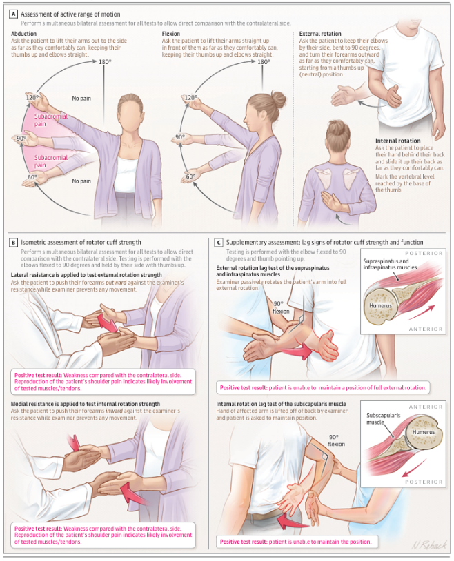
Examination: For most the diagnosis can be suspected and confirmed by a detailed history and clinical exam. Active range of motion (abduction, flexion, internal/external rotation) should be compared bilaterally; a painful (active) range of motion (ROM) with preserved passive motion suggests subacromial pain, while global restriction of both active and passive motion points to adhesive capsulitis or osteoarthritis, in most. Resisted strength testing identifies weakness from pain inhibition, cuff tears, or neurologic causes. Lag signs, external rotation lag (supraspinatus/infraspinatus) and internal rotation lag (subscapularis), supplement resisted testing and are particularly useful because subscapularis weakness can be masked by pectoralis/latissimus substitution in neutral position. Importantly, the classic "special tests" (Neer, Hawkins-Kennedy, Speed, empty can) have low interrater reliability and cannot reliably localize a structural cause; they should not drive diagnosis or imaging decisions.
Imaging. Routine imaging is not recommended for most new shoulder pain, regardless of symptom duration, because structural abnormalities (cuff tears, calcific deposits, labral changes) are common in asymptomatic shoulders and increase with age. A large population-based study of 602 participants confirmed this overlap. This aligns with other reports (recent RheumNow commentary) that rotator cuff abnormalities are very common on MRI in patients over 40. Expensive imaging may yield incidental, clinically irrelevant findings that risk overdiagnosis, anxiety, and unnecessary intervention. Imaging is appropriate only for suspected infection, malignancy, major trauma (fracture/dislocation), significant or progressive neurologic weakness, or symptoms that worsen rather than plateau. When imaging is indicated, start with plain radiographs; ultrasonography and MRI have comparable accuracy for full-thickness tears, with MRI preferred when detailed soft-tissue planning is needed.
Treatment: What is evidence-based and guideline-endorsed?
High-certainty evidence: education, activity modification, and watchful waiting (favorable natural history); landmark-guided glucocorticoid injection for moderate-severe pain (no added benefit from imaging guidance); topical NSAIDs as first-line pharmacotherapy.
Guideline-endorsed but modest-benefit: supervised exercise (small, clinically unimportant benefit over placebo per Cochrane data, though a single physical therapy session performs as well as a 6-session program); oral NSAIDs (trivial-to-small benefit, weigh against GI/renal/cardiovascular risk).
Not recommended: subacromial decompression surgery (high-certainty evidence of no benefit over placebo surgery); hyaluronic acid and platelet-rich plasma injections (no meaningful benefit); opioids; extracorporeal shockwave therapy; manual therapy alone.
Surgery for full-thickness rotator cuff tears remains of uncertain benefit versus nonoperative care in patients over 55 with small, degenerative tears — shared decision-making is essential.
Continue Reading
Related Content



ADD THE FIRST COMMENT
Disclosures
Disclosures
The author has no conflicts of interest to disclose related to this subject
The author used AI to research and organize this content, and maintains responsibility for its accuracy



If you are a health practitioner, you may Login/Register to comment.
Due to the nature of these comment forums, only health practitioners are allowed to comment at this time.